. 2020 Sep 21;10(1):15360.
doi: 10.1038/s41598-020-72730-z.

DNA methylation across the genome in aged human skeletal muscle tissue and muscle-derived cells: the role of HOX genes and physical activity

Affiliations

DNA methylation across the genome in aged human skeletal muscle tissue and muscle-derived cells: the role of HOX genes and physical activity

D C Turner et al. Sci Rep. .

Abstract

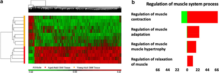
Skeletal muscle tissue demonstrates global hypermethylation with age. However, methylome changes across the time-course of differentiation in aged human muscle derived cells, and larger coverage arrays in aged muscle tissue have not been undertaken. Using 850K DNA methylation arrays we compared the methylomes of young (27 ± 4.4 years) and aged (83 ± 4 years) human skeletal muscle and that of young/aged heterogenous muscle-derived human primary cells (HDMCs) over several time points of differentiation (0, 72 h, 7, 10 days). Aged muscle tissue was hypermethylated compared with young tissue, enriched for; pathways-in-cancer (including; focal adhesion, MAPK signaling, PI3K-Akt-mTOR signaling, p53 signaling, Jak-STAT signaling, TGF-beta and notch signaling), rap1-signaling, axon-guidance and hippo-signalling. Aged cells also demonstrated a hypermethylated profile in pathways; axon-guidance, adherens-junction and calcium-signaling, particularly at later timepoints of myotube formation, corresponding with reduced morphological differentiation and reductions in MyoD/Myogenin gene expression compared with young cells. While young cells showed little alterations in DNA methylation during differentiation, aged cells demonstrated extensive and significantly altered DNA methylation, particularly at 7 days of differentiation and most notably in focal adhesion and PI3K-AKT signalling pathways. While the methylomes were vastly different between muscle tissue and HDMCs, we identified a small number of CpG sites showing a hypermethylated state with age, in both muscle tissue and cells on genes KIF15, DYRK2, FHL2, MRPS33, ABCA17P. Most notably, differential methylation analysis of chromosomal regions identified three locations containing enrichment of 6-8 CpGs in the HOX family of genes altered with age. With HOXD10, HOXD9, HOXD8, HOXA3, HOXC9, HOXB1, HOXB3, HOXC-AS2 and HOXC10 all hypermethylated in aged tissue. In aged cells the same HOX genes (and additionally HOXC-AS3) displayed the most variable methylation at 7 days of differentiation versus young cells, with HOXD8, HOXC9, HOXB1 and HOXC-AS3 hypermethylated and HOXC10 and HOXC-AS2 hypomethylated. We also determined that there was an inverse relationship between DNA methylation and gene expression for HOXB1, HOXA3 and HOXC-AS3. Finally, increased physical activity in young adults was associated with oppositely regulating HOXB1 and HOXA3 methylation compared with age. Overall, we demonstrate that a considerable number of HOX genes are differentially epigenetically regulated in aged human skeletal muscle and HDMCs and increased physical activity may help prevent age-related epigenetic changes in these HOX genes.


Conflict of interest statement

The authors declare no competing interests.

Figures

Figure 1
Figure 1
DNA methylation across the genome in aged human skeletal muscle tissue compared with young adult tissue. (a) Hierarchical clustering heatmap of the significantly differentially methylated CpG sites, depicting aged skeletal muscle containing a hypermethylated (RED) versus hypomethylated (GREEN) signature compared with young adult tissue. (b) CpG sites for most significantly enriched GO term containing the search term ‘muscle’; ‘regulation of muscle system process’ (RED hypermethylated and GREEN hypomethylated CpGs). X axis labels = number of DMPs.
Figure 2
Figure 2
Aged and young adult HDMCs differentiated over 0, 72 h, 7d and 10 d and differences in DNA methylation. (a) Light microscope images of aged versus young HDMCs, depict fewer myotubes in aged versus young cells, particularly at 7 and 10 days. (b) MyoD and myogenin gene expression in aged versus young cells over the differentiation time course. Where, reductions in myogenin were observed at 72 h in aged cells as well as a delayed increase in the upregulation of myogenin compared with young cells. (c) Hierarchical clustering heatmap of significant DMPs between aged and young HDMCs across the entire time-course of differentiation (all time points of 0, 72 h, 7 d and 10 d). RED hypermethylated and GREEN hypomethylated. (d) Comparison of DNA methylation in aged versus young HDMCs at 7 d of differentiation (7 d aged vs. 7 d young adult cells) in most enriched KEGG pathway, ‘focal adhesion’. Copyright permission for KEGG pathway image obtained from Kanehisa Laboratories. Note, the most significant (lowest p-value) DMP for each gene is used to colour this pathway image. Therefore, this is not always accurate where multiple DMPs occur for a single gene and the image is therefore only a visual representation of the overarching methylation profile in this pathway. Full and accurate DMP lists for 'focal adhesion' pathway in these conditions can be found in  Suppl. File 5i.
Figure 3
Figure 3
SOM profiling of DNA methylation over the time-course of differentiation in aged versus young adult HDMCs. (a) Demonstrates a larger number of hypomethylated and hypermethylated CpG sites in aged HDMCs, particularly at 7 days of differentiation, compared with young adult HDMCs. (b) Venn Diagram analysis depicting the 24 common CpG sites that were altered at every time point of differentiation between aged and young adult muscle HDMCs cells (0, 72 h, 7d and 10 d). c. As with the above analysis in 3a, SOM profiling identified that 16 out of these 24 CpG’s also demonstrated the most alterations in methylation at 7 days of differentiation in aged cells.
Figure 4
Figure 4
Venn diagram of common overlapping significant DMPs between aged skeletal muscle tissue and HDMCs. Overlap of the tissue and aged cells identified 6 common DMPs on genes KIF15 (2 CpG’s Cg00702638 & cg24888989), DYRK2 (cg09516963), FHL2 (cg22454769), MRPS33 (cg26792755) and ABCA17P (cg02331561). Given that aged cells demonstrated the majority of changes in methylation at 7 days versus young cells. When overlapping the 7 d most significantly differentially methylated CpG lists, 4 DMPs (out of the 6 DMPs identified above) were also identified, including: MGC87042 (cg00394316), C2orf70 (cg23482427), ABCA17P (cg02331561) and cg27209395 (not an annotated gene). Once more, all of these DMPs (with the exception of C2orf70, cg23482427) were hypermethylated in the tissue analysis as well as the HDMCs. With ABCA17P (cg02331561) highlighted across all DMP lists.
Figure 5
Figure 5
HOX family of genes and their DNA methylation in aged tissue and HDMCs. (a) Venn diagram analysis identifying 9 overlapping differentially methylated HOX genes: including HOXD10, HOXD9, HOXD8, HOXA3, HOXC9, HOXB1, HOXB3, HOXC-AS2 and HOXC10 (note this Venn diagram analysis is by ‘gene symbol’ not ‘probe cg’ (CpG site), as some HOX genes also had more than 1 CpG per gene symbol, full CpG lists are located in Suppl. Figure 9a,b,c,d). (b) All HOX family genes by CpG site (cg probe) differentially methylated in aged compared with young skeletal muscle tissue, predominantly all demonstrating hypermethylation. (c) SOM profiling depicting the temporal regulation of DNA methylation in aged HDMCs as they differentiate in CpGs located amongst the HOX family of genes (depicting the 9 HOX genes altered, by gene symbol, in both the tissue and cells). The majority of these HOX CpG sites were differentially methylated at 7 days of differentiation in the aged cells.
Figure 6
Figure 6
Gene expression of the HOX family of genes in aged compared to young HDMCs at 7 days of differentiation.
Figure 7
Figure 7
Schematic of the overarching main results. Hypermethylation of the genome and the HOX genes in aging skeletal muscle tissue (RED box) compared with young adult tissue (GREEN BOX). Dysregulation of HOX genes in aged cells particularly at 7 days of differentiation in muscle derived cells. Inverse methylation (hypermethylation- red circles) with gene expression (reduced- red rectangle) in aging cells particularly for HOXB1 and HOXA3. Increased physical activity levels are associated with the hypomethylation (fewer red circles more white circles) of the same two genes, HOXB1 and HOXA3.

Similar articles

Cited by

References

    1. Sharples AP, Hughes DC, Deane CS, Saini A, Selman C, Stewart CE. Longevity and skeletal muscle mass: the role of IGF signalling, the sirtuins, dietary restriction and protein intake. Aging Cell. 2015;14:511–523. - PMC - PubMed
    1. Morley JE, Anker SD, von Haehling S. Prevalence, incidence, and clinical impact of sarcopenia: facts, numbers, and epidemiology-update 2014. J. Cachexia Sarcopenia Muscle. 2014;5:253–259. - PMC - PubMed
    1. Shafiee G, Keshtkar A, Soltani A, Ahadi Z, Larijani B, Heshmat R. Prevalence of sarcopenia in the world: a systematic review and meta- analysis of general population studies. J. Diabetes Metab. Disord. 2017;16:21. - PMC - PubMed
    1. Hughes VA, et al. Longitudinal muscle strength changes in older adults: influence of muscle mass, physical activity, and health. J. Gerontol. Ser. A-Biol. Sci. Med. Sci. 2001;56:B209–217. - PubMed
    1. Cruz-Jentoft AJ, et al. Sarcopenia: European consensus on definition and diagnosis: report of the European Working Group on Sarcopenia in Older People. Age Ageing. 2010;39:412–423. - PMC - PubMed

Publication types