. 2020 Sep 21;10(1):15402.
doi: 10.1038/s41598-020-72373-0.

Structure and function of bacteriophage CBA120 ORF211 (TSP2), the determinant of phage specificity towards E. coli O157:H7

Affiliations

Structure and function of bacteriophage CBA120 ORF211 (TSP2), the determinant of phage specificity towards E. coli O157:H7

Julia Greenfield et al. Sci Rep. .

Abstract

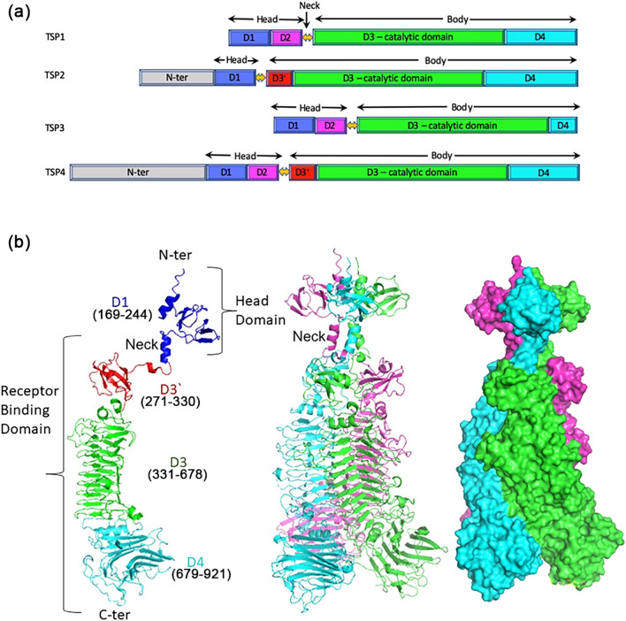
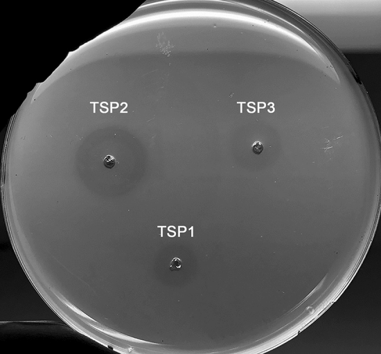
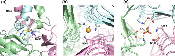
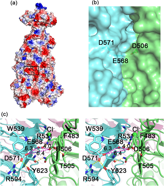
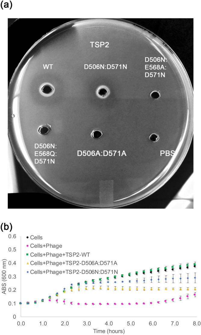
The genome of Escherichia coli O157:H7 bacteriophage vB_EcoM_CBA120 encodes four distinct tailspike proteins (TSPs). The four TSPs, TSP1-4, attach to the phage baseplate forming a branched structure. We report the 1.9 Å resolution crystal structure of TSP2 (ORF211), the TSP that confers phage specificity towards E. coli O157:H7. The structure shows that the N-terminal 168 residues involved in TSPs complex assembly are disordered in the absence of partner proteins. The ensuing head domain contains only the first of two fold modules seen in other phage vB_EcoM_CBA120 TSPs. The catalytic site resides in a cleft at the interface between adjacent trimer subunits, where Asp506, Glu568, and Asp571 are located in close proximity. Replacement of Asp506 and Asp571 for alanine residues abolishes enzyme activity, thus identifying the acid/base catalytic machinery. However, activity remains intact when Asp506 and Asp571 are mutated into asparagine residues. Analysis of additional site-directed mutants in the background of the D506N:D571N mutant suggests engagement of an alternative catalytic apparatus comprising Glu568 and Tyr623. Finally, we demonstrate the catalytic role of two interacting glutamate residues of TSP1, located in a cleft between two trimer subunits, Glu456 and Glu483, underscoring the diversity of the catalytic apparatus employed by phage vB_EcoM_CBA120 TSPs.


Conflict of interest statement

The authors declare no competing interests.

Figures

Figure 1
Figure 1
Domain structures of CBA120 TSPs and overall structure of TSP2. (a) Schematic diagram of TSP1-4 domain arrangements. The N-terminal regions (N-ter) of TSP2 and TSP4 contain 2 and 4 domains, respectively. These domains are involved in protein–protein interactions and are not depicted here because the TSP2 N-ter region (residues 1–168) is structurally disordered. (b) A cartoon representation of the monomer (left), the trimer (middle), and the trimer surface representation of TSP2 (right). Each molecule of the trimer assembly is colored differently. The figure was generated using the computer program PyMol v1.8.0.2 (Schrödinger, LLC, https://www.pymol.org).
Figure 2
Figure 2
Halo assay of TSP1, TSP2, and TSP3. E. coli strain ATCC 700,728 was embedded in agarose. Wells (3 mm) were cut out of the agarose and loaded with 10 µL (6 mg/mL) of TSPs. Following overnight incubation at 37 °C, each TSP produced a halo, which is indicative of glycosidase activity.
Figure 3
Figure 3
Phage infection assays. Phage CBA120 infection of E. coli strain ATCC 700,728 was followed by spectrophotometry at 600 nm as detailed in the methods section. The mean values of technical triplicate and the standard deviations are shown (a) Phage infection in the presence of various TSPs. The ability of CBA120 to infect E. coli O157:H7 treated with TSP1 (yellow circles), TSP2 (green circles), or TSP3 (blue circles) at 100 μg/mL is shown together with controls consisting of bacterial cells alone (black circles) and non-TSP treated bacterial cells incubated with phage (magenta circles). Only treatment with TSP2 prevented phage infection and subsequent bacterial death. (b) Phage CBA120 infection dependence on TSP2 concentration. Controls are the same as in (a). TSP2 at 25 and 100 μg/mL prevented killing by the phage but 5 μg/mL inhibited phage killing only for ~ 4.5 h.
Figure 4
Figure 4
TSP2 D1 and D3′ folds. (a) Cartoon representation of D1 with rainbow coloring from the N-terminus (blue) to the C-terminus (red). (b) Stereoscopic view of superimposed D1 fold modules of TSP2 (red; residues q66-247), TSP4 (gray; residues 340–421 PDB entry 5W6H)), TSP1 (green; residues 12–96, PDB entry code 4OJ5), TSP3 (light blue; residues 12–95, PDB entry 6NW9), and TSP gp63.1 from E. coli 4 s G7C phage (cyan; residues 13–96, PDB entry 4QNL). (c) Cartoon representation of the D3′ with rainbow coloring from the N-terminus (blue) to the C-terminus (red). (d) Stereoscopic view of superimposed D3′ of TSP2 (red; residues 270–328), TSP4 (gray; residues 503–557 PDB entry 5W6H), TSP gp49 from A. baumannii phage Fri1 (green; residues 209–269, PDB entry 6C72), TSP from A. baumannii phage AM24 (light blue; residues 37–96, PDB entry 5W5P), TSP gp42 from A. baumannii phage vb_AbaP_AS12 (cyan; residues 23–82, PDB entry 6EU4). The figure was generated using the computer program PyMol v1.8.0.2 (Schrödinger, LLC, https://www.pymol.org).
Figure 5
Figure 5
Stereoscopic representation of the TSP2 D3 and D4 folds. The polypeptide chain is colored with a rainbow color scheme beginning with blue at the N-terminus and ending in red at the C-terminus of the entire molecule. (a) The D3 β-helix, indicating the two capping α-helices. (b) The D4 β-sandwich. The D4 α-helix is close to the C-terminus of the polypeptide chain and caps the D3 β-helix C-terminus, a feature unique to TSP2. The figure was generated using the computer program PyMol v1.8.0.2 (Schrödinger, LLC, https://www.pymol.org).
Figure 6
Figure 6
Anions located along the TSP2 threefold axis support trimer oligomerization. (a) A chloride located between the neck and the D3′ domain forms charge-charge interactions with three guanidinium groups of Arg284. (b) A chloride located along the intermolecular D3 β-helices axis interacts with three side chain amide groups of Asn565. (c) A sulfate located along the intermolecular D3 β-helices axis form charge-charge interaction with the carboxyl groups of Asp415 and the amino groups of Lys465. The figure was generated using the computer program PyMol v1.8.0.2 (Schrödinger, LLC, https://www.pymol.org).
Figure 7
Figure 7
The active site of TSP2. (a) Surface vacuum electrostatics of the TSP2 trimer, indicating the location of the active site (rectangular black box). Negatively polar regions are colored red and positively polar region are colored blue. The overall polarity of the active site is negative. (b) The partially transparent molecular surface at the active site, highlighting key active site residues. As can be seen, the interface between the green and cyan molecules forms a cleft. The trimer contains three such clefts at the three subunit interfaces. (c) Stereoscopic representation of active site residues. The distances between the three carboxyl groups of Asp506, Asp571, and Glu578, the candidate catalytic resides that were probed by site-directed mutagenesis, are shown in magenta dash lines. Key salt bridges and hydrogen bond interactions are shown in yellow dash lines. Aromatic groups may interact with the substrate pyranose rings. The chloride bound underneath the active site is shown as green sphere. The trimer subunits are colored differently. The figure was generated using the computer program PyMol v1.8.0.2 (Schrödinger, LLC, https://www.pymol.org).
Figure 8
Figure 8
Assays to probe TSP2 active site mutants. Structure and mechanism-based analyses were used to identify the active site residues. (a) In the halo assay, E. coli strain ATCC 700,728 was embedded in agarose. Wells (3 mm) were cut out of the agarose and loaded with 10 μL (6 mg/mL) of wild-type TSP2 or active site TSP2 mutants, and incubated overnight at 37 °C to visualize glycosidase activity. The D506A:D571A TSP2 mutant was incapable of producing a halo, indicating glycosidase activity of TSP2 was abolished. Conversely, the D506:D571N TSP2 mutant retained enzymatic activity. However, introducing the E568A (i.e. D506N:E568A:D571N) or E568Q (i.e. D506N:D568Q:D571N) mutations to a D506N:D571N background inhibited TSP2 glycosidase activity, suggesting involvement of the Glu568 carboxyl group as part of an alternative catalytic machinery. Wild-type TSP2 and PBS only served as positive and negative controls for glycosidase activity, respectively. (b) In the bacterial infection assay, E. coli O157:H7 (ATCC 700728) culture was mixed with TSP variants as described in Methods. Absorbance measurements at 600 nm were made every 20 min. Bacterial growth is shown for E. coli incubated with phage following treatment with 100 μg/mL of either wild-type or TSP2 (green squares, D506:D571A TSP2 (yellow triangles), or D506N:D571N TSP2 (blue diamonds). Growth of bacterial cells alone (black circles) and non-TSP treated bacterial cells incubated with phage (magenta circles) served as controls. The mean values of technical triplicate and the standard deviations are shown.
Figure 9
Figure 9
TSP1 active site structure and halo assay of active site residues. Structure and mechanism-based analyses were used to identify the active site residues. (a) Depiction of key carboxylic acid residues in the interface between two TSP1 subunits. The figure was generated using the computer program PyMol v1.8.0.2 (Schrödinger, LLC, https://www.pymol.org). (b) Halo assay of wild-type and mutant TSP1. E. coli strain ATCC 700728 was embedded in agarose. Wells (3 mm) were cut out of the agarose and loaded with 10 µL (6 mg/mL) of active site TSP1 mutants, and incubated overnight at 37 °C to visualize glycosidase activity. The absence of a halo for E456A:E483A TSP1 suggests an inhibition of enzymatic activity. Alternatively, the appearance of a halo for the E456Q:E483Q mutant indicates TSP1 retains the ability to display glycosidase activity. Wild-type TSP1 and PBS only served as positive and negative controls for glycosidase activity, respectively.

References

    1. King AMQ, et al. Changes to taxonomy and the international code of virus classification and nomenclature ratified by the international committee on taxonomy of viruses (2018) Arch. Virol. 2018 doi: 10.1007/s00705-018-3847-1. - DOI - PubMed
    1. Park M, et al. Characterization and comparative genomic analysis of a novel bacteriophage, SFP10, simultaneously inhibiting both Salmonella enterica and Escherichia coli O157:H7. Appl. Environ. Microbiol. 2012;78:58–69. doi: 10.1128/AEM.06231-11. - DOI - PMC - PubMed
    1. Bavaro MFE. coli O157:H7 and other toxigenic strains: the curse of global food distribution. Curr. Gastroenterol. Rep. 2012;14:317–323. doi: 10.1007/s11894-012-0264-6. - DOI - PubMed
    1. Kutter EM, et al. Characterization of a ViI-like phage specific to Escherichia coli O157:H7. Virol. J. 2011;8:430. doi: 10.1186/1743-422X-8-430. - DOI - PMC - PubMed
    1. Lim JY, Yoon JW, Hovde CJ. A brief overview of Escherichia coli O157:H7 and its plasmid O157. J. Microbiol. Biotechnol. 2010;20:5–14. - PMC - PubMed

Publication types

MeSH terms

Substances

LinkOut - more resources