

Sagittal knee kinematics in relation with the posterior tibia slope during jump landing after an anterior cruciate ligament reconstruction
- PMID: 32959098
- PMCID: PMC7505908
- DOI: 10.1186/s40634-020-00289-9
Sagittal knee kinematics in relation with the posterior tibia slope during jump landing after an anterior cruciate ligament reconstruction
Abstract
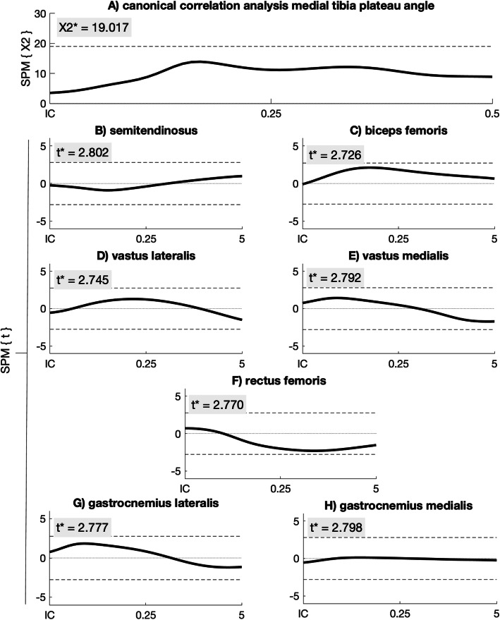
Purpose: An increased posterior tibia plateau angle is associated with increased risk for anterior cruciate ligament injury and re-rupture after reconstruction. The aims of this study were to determine whether the tibia plateau angle correlates with dynamic anterior tibia translation (ATT) after an anterior cruciate ligament reconstruction and whether the tibia plateau angle correlates with aspects of knee kinematics and kinetics during jump landing.
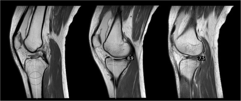
Methods: Thirty-seven patients after anterior cruciate ligament reconstruction with autograft hamstring tendon were included. Knee flexion angle and knee extension moment during single leg hops for distance were determined using a motion capture system and the dynamic ATT with its embedded method. The medial and lateral posterior tibia plateau angle were measured using MRI. Moreover, passive ATT was measured using the KT-1000 arthrometer.
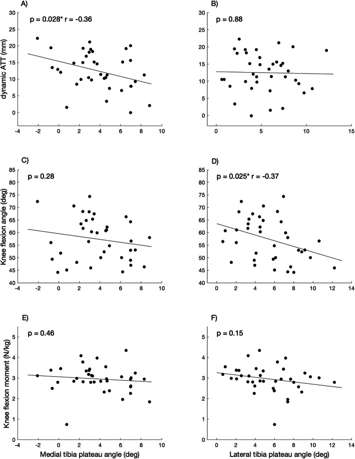
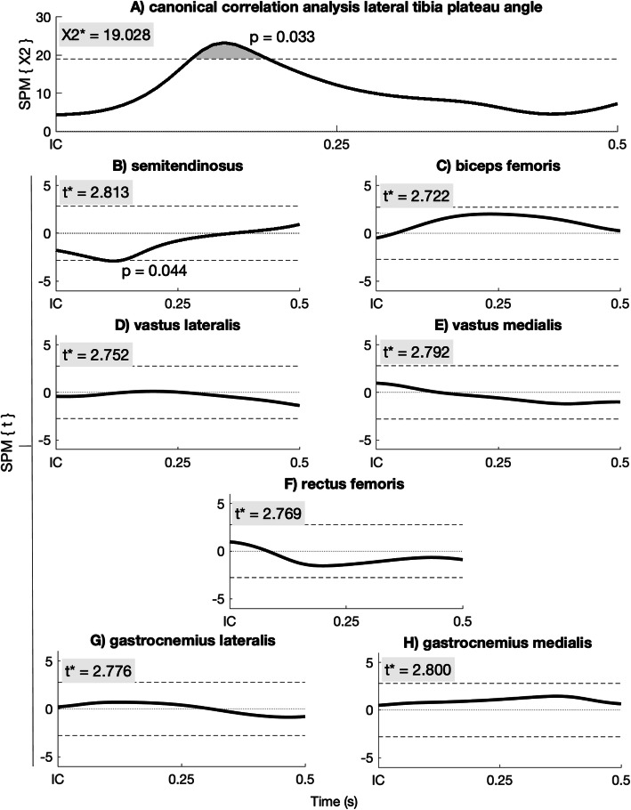
Results: A weak negative correlation was found between the maximal dynamic ATT and the medial tibia plateau angle (p = 0.028, r = - 0.36) and between the maximal knee flexion angle and the lateral tibia plateau angle (p = 0.025, r = - 0.37) during landing. Patients with a smaller lateral tibia plateau angle show larger maximal knee flexion angle during landing than the patients with larger lateral tibia plateau angle. Also, the lateral tibia plateau angle is associated the amount of with muscle activity.
Conclusion: The posterior medical tibia plateau angle is associated with dynamic ATT. The maximal knee flexion angle and muscle activity are associated with the posterior lateral tibia plateau angle.
Level of evidence: III.
Keywords: Anatomy; In-vivo knee kinematics; Knee; Tibia plateau.
Conflict of interest statement
The authors declare that they have no conflict of interest.
Figures
Similar articles
-
Healthy subjects with lax knees use less knee flexion rather than muscle control to limit anterior tibia translation during landing.J Exp Orthop. 2020 May 15;7(1):32. doi: 10.1186/s40634-020-00246-6. J Exp Orthop. 2020. PMID: 32415565 Free PMC article.
-
Variations in Knee Kinematics After ACL Injury and After Reconstruction Are Correlated With Bone Shape Differences.Clin Orthop Relat Res. 2017 Oct;475(10):2427-2435. doi: 10.1007/s11999-017-5368-8. Clin Orthop Relat Res. 2017. PMID: 28451863 Free PMC article.
-
Posterior tibial slope influences static anterior tibial translation in anterior cruciate ligament reconstruction: a minimum 2-year follow-up study.Am J Sports Med. 2014 Apr;42(4):927-33. doi: 10.1177/0363546514521770. Epub 2014 Feb 19. Am J Sports Med. 2014. PMID: 24553814
-
Effects of Anterior Closing Wedge Tibial Osteotomy on Anterior Cruciate Ligament Force and Knee Kinematics.Am J Sports Med. 2018 Feb;46(2):370-377. doi: 10.1177/0363546517736767. Epub 2017 Nov 3. Am J Sports Med. 2018. PMID: 29100001
-
Passive anterior tibia translation in anterior cruciate ligament-injured, anterior cruciate ligament-reconstructed and healthy knees: a systematic review.Musculoskelet Surg. 2019 Aug;103(2):121-130. doi: 10.1007/s12306-018-0572-6. Epub 2018 Oct 16. Musculoskelet Surg. 2019. PMID: 30328030 Free PMC article.
Cited by
-
Lower anatomical femoral ACL tunnel can be created in the large volume of femoral intercondylar notch.Knee Surg Sports Traumatol Arthrosc. 2022 Oct;30(10):3322-3327. doi: 10.1007/s00167-022-06921-8. Epub 2022 Feb 24. Knee Surg Sports Traumatol Arthrosc. 2022. PMID: 35201373
-
Posterior tibial slope and meniscal slope correlate with in vivo tibial internal rotation during running and drop jump.Knee Surg Sports Traumatol Arthrosc. 2023 Jun;31(6):2366-2373. doi: 10.1007/s00167-022-07163-4. Epub 2022 Sep 17. Knee Surg Sports Traumatol Arthrosc. 2023. PMID: 36115904
-
Knees with straight Blumensaat's line have small volume of femoral intercondylar notch.Knee Surg Sports Traumatol Arthrosc. 2022 Jan;30(1):102-108. doi: 10.1007/s00167-021-06677-7. Epub 2021 Jul 20. Knee Surg Sports Traumatol Arthrosc. 2022. PMID: 34283249
References
-
- Boeth H, Duda GN, Heller MO, Ehrig RM, Doyscher R, Jung T, Moewis P, Scheffler S, Taylor WR. Anterior cruciate ligament-deficient patients with passive knee joint laxity have a decreased range of anterior-posterior motion during active movements. Am J Sports Med. 2013;41:1051–1057. doi: 10.1177/0363546513480465. - DOI - PubMed
-
- Dejour D, Pungitore M, Valluy J, Nover L, Saffarini M, Demey G (2019) Tibial slope and medial meniscectomy significantly influence short-term knee laxity following ACL reconstruction. Knee Surg Sports Traumatol Arthrosc. 27(11):3481-3489 - PubMed
LinkOut - more resources
Full Text Sources
Research Materials
