

Expression of Mucins in Salivary Gland Mucoepidermoid Carcinoma
- PMID: 32959209
- PMCID: PMC8134608
- DOI: 10.1007/s12105-020-01226-z
Expression of Mucins in Salivary Gland Mucoepidermoid Carcinoma
Abstract
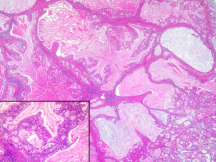
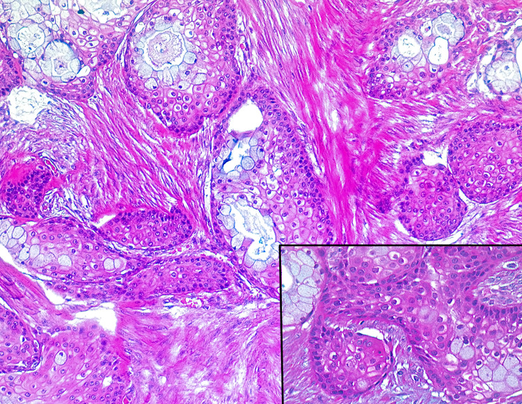
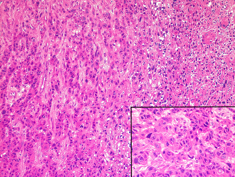
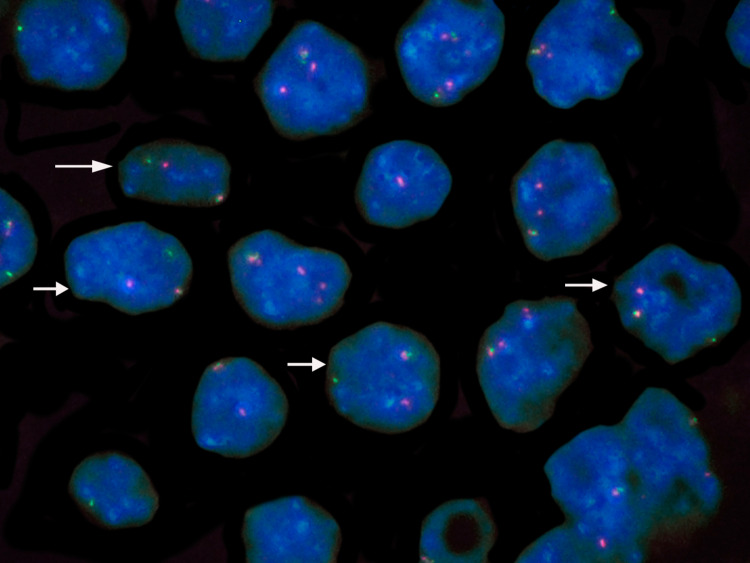
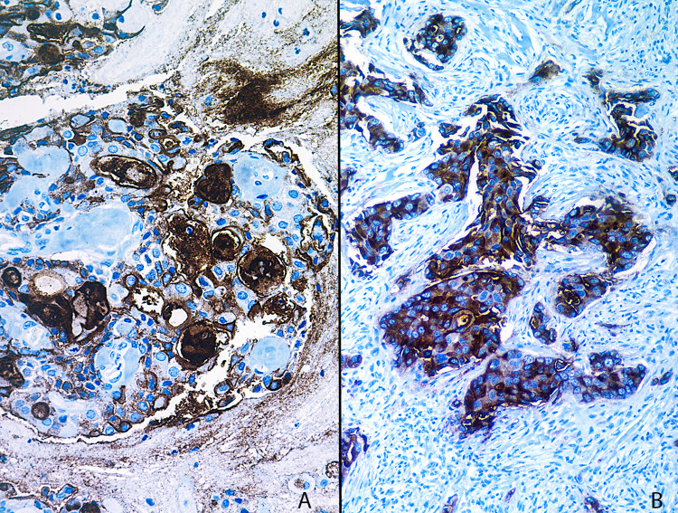
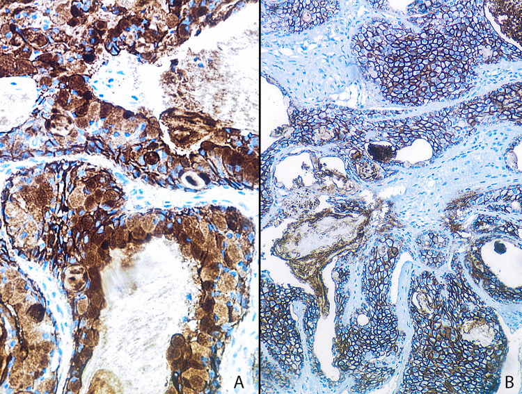
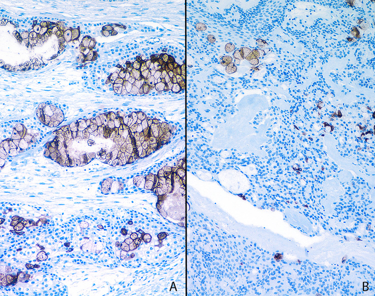
Mucoepidermoid carcinoma (MEC) is the most common malignant salivary gland tumour in both adults and children. Histological grading of MEC is subjective, but plays an important role in predicting patient prognosis. Epithelial mucin (MUC) status may aid in establishing a more accurate grade. This study aimed to investigate the expression of various mucins (MUC1, MUC2, MUC4 and MUC5AC) in MECs to determine a possible correlation with tumour grade. Fifteen cases of each tumour grade (low-, intermediate-, and high-grade) were retrieved from the pathology archives of the Department of Oral Pathology and Oral Biology at the University of Pretoria. The patients included 23 men and 22 women, and ranged from 13 to 85 years (mean 49.8 years). Sections from formalin-fixed paraffin-embedded (FFPE) tissue were used for fluorescence in situ hybridization (FISH) for MAML2 rearrangements and MUC immunohistochemical analysis. The percentage immunohistochemical expression of the neoplastic mucous cells was evaluated first, followed by the overall percentage expression of all tumour cells. The results indicated that MUC1 overexpression may be a reliable marker of high-grade MECs, whereas MUC4 overexpression may be more indicative of low-grade tumours. MUC5AC expression was considered an unreliable marker in determining grade. MUC2 was only expressed in a single case of MEC and may be considered a useful marker to exclude MEC as a diagnostic possibility. This study demonstrates that MECs show an altered MUC expression pattern that can be used for diagnostic purposes and to aid in establishing a more accurate tumour grade.
Keywords: Epithelial mucin expression; FISH; Histological grade; Immunohistochemistry; Mucoepidermoid carcinoma; Salivary gland carcinoma.
Conflict of interest statement
The authors declare that they have no conflict of interest.
Figures
Similar articles
-
MUC1, MUC2, MUC4, and MUC5AC expression in salivary gland mucoepidermoid carcinoma: diagnostic and prognostic implications.Am J Surg Pathol. 2005 Jul;29(7):881-9. doi: 10.1097/01.pas.0000159103.95360.e8. Am J Surg Pathol. 2005. PMID: 15958852
-
Expression of membrane-bound mucins (MUC1 and MUC4) and secreted mucins (MUC2, MUC5AC, MUC5B, MUC6 and MUC7) in mucoepidermoid carcinomas of salivary glands.Am J Surg Pathol. 2005 Jun;29(6):806-13. doi: 10.1097/01.pas.0000155856.84553.c9. Am J Surg Pathol. 2005. PMID: 15897748
-
"Pancreatic Mucoepidermoid Carcinoma" Is not a Pancreatic Counterpart of CRTC1/3-MAML2 Fusion Gene-related Mucoepidermoid Carcinoma of the Salivary Gland, and May More Appropriately be Termed Pancreatic Adenosquamous Carcinoma With Mucoepidermoid Carcinoma-like Features.Am J Surg Pathol. 2018 Nov;42(11):1419-1428. doi: 10.1097/PAS.0000000000001135. Am J Surg Pathol. 2018. PMID: 30138216
-
Mucoepidermoid carcinoma of minor salivary glands: a clinical study of 16 cases and review of the literature.Oral Dis. 2006 Jul;12(4):364-70. doi: 10.1111/j.1601-0825.2005.01166.x. Oral Dis. 2006. PMID: 16792720 Review.
-
Recent advances in mucin immunohistochemistry in salivary gland tumors and head and neck squamous cell carcinoma.Oral Oncol. 2011 Sep;47(9):797-803. doi: 10.1016/j.oraloncology.2011.06.008. Epub 2011 Jul 1. Oral Oncol. 2011. PMID: 21723776 Review.
Cited by
-
Central mucoepidermoid carcinoma of the mandible deriving from odontogenic cyst: A case report and review of the literature.Clin Case Rep. 2022 Aug 3;10(8):e6040. doi: 10.1002/ccr3.6040. eCollection 2022 Aug. Clin Case Rep. 2022. PMID: 35937015 Free PMC article.
-
Metastatic cutaneous squamous cell carcinoma accounts for nearly all squamous cell carcinomas of the parotid gland.Virchows Arch. 2024 Jul;485(1):3-11. doi: 10.1007/s00428-024-03798-5. Epub 2024 Apr 17. Virchows Arch. 2024. PMID: 38630141 Free PMC article. Review.
-
Clinicopathological Analysis of Salivary Glands Warthin-like Mucoepidermoid Carcinoma: A Systematic Review.Head Neck Pathol. 2025 May 8;19(1):55. doi: 10.1007/s12105-025-01778-y. Head Neck Pathol. 2025. PMID: 40338433
-
Preparation of Soluble Mucin Solutions from the Salivary Glands.Methods Mol Biol. 2024;2763:45-50. doi: 10.1007/978-1-0716-3670-1_3. Methods Mol Biol. 2024. PMID: 38347398
-
Expression of MUC1, MUC2 and MUC5AC in salivary gland mucoepidermoid carcinoma: A case series with diagnostic implications.J Oral Maxillofac Pathol. 2022 Jul-Sep;26(3):346-351. doi: 10.4103/jomfp.jomfp_342_21. Epub 2022 Oct 17. J Oral Maxillofac Pathol. 2022. PMID: 36588842 Free PMC article.
References
-
- Ellis GL, Auclair PL. AFIP atlas of tumor pathology. Washington, DC: American Registry of Pathology in Collaboration with the Armed Forces Institute of Pathology; 2008.
-
- El-Naggar AK, Chan JKC, Rubin-Grandis J, Takata T, Slootweg PJ, International Agency for Research on Cancer . World Health Organization classification of tumours. 4. Lyon: International Agency for Research on Cancer; 2017.
-
- Griffith CC, Seethala RR. Mucoepidermoid carcinoma of minor salivary glands: potential pitfalls and prognostic challenges. Pathol Case Rev. 2011;16(4):145–150. doi: 10.1097/PCR.0b013e31822489f8. - DOI
MeSH terms
Substances
LinkOut - more resources
Full Text Sources
Medical
Research Materials
Miscellaneous
