

Primary Intraosseous Adenoid Cystic Carcinoma with Widespread Skeletal Metastases Showing Features of High-Grade Transformation
- PMID: 32959213
- PMCID: PMC8134797
- DOI: 10.1007/s12105-020-01228-x
Primary Intraosseous Adenoid Cystic Carcinoma with Widespread Skeletal Metastases Showing Features of High-Grade Transformation
Abstract
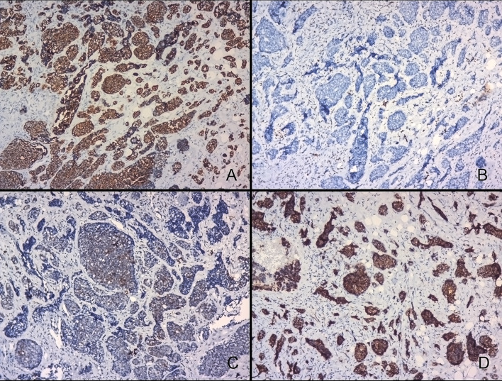
Malignant salivary gland carcinomas arising primarily within the jaw bones are extremely rare. The most common salivary malignancy in these locations is mucoepidermoid carcinoma followed by adenoid cystic carcinoma and adenocarcinoma. The clinical picture and imaging studies of these malignancies may be confused with odontogenic lesions which are more common in this location. Adenoid cystic carcinomas have a prolonged clinical course, tendency for perineural invasion and distant metastasis and multiple recurrences. The diagnosis of these tumors requires thorough histopathologic examination. Immunohistochemical studies may be required in cases showing solid growth pattern. High-grade transformation, earlier termed as dedifferentiation, has been observed in several salivary gland carcinomas including adenoid cystic carcinoma. These transformed tumors are reported to have an extremely poor prognosis. Here, we report a case of primary intraosseous adenoid cystic carcinoma with extensive skeletal metastases which showed a negative staining with p63 and positive staining with CD117. The tumor had a predominant solid growth pattern with areas indicative of high-grade transformation. A negative p63 staining may indicate an incomplete or focal loss of abluminal layer and this is one of the criteria for high-grade transformation in adenoid cystic carcinoma.
Keywords: Adenoid cystic carcinoma; Female; Head and neck neoplasms; Humans; Immunohistochemistry; Mandible; Paraesthesia; Salivary gland neoplasms.
Conflict of interest statement
All authors declare that they have no conflict of interest to disclose.
Figures


Similar articles
-
High-grade transformation/dedifferentiation of an adenoid cystic carcinoma of the minor salivary gland to myoepithelial carcinoma.Pathol Int. 2018 Feb;68(2):133-138. doi: 10.1111/pin.12624. Epub 2017 Dec 29. Pathol Int. 2018. PMID: 29287310
-
Bilateral intraosseous adenoid cystic carcinoma of the mandible: report of a case with lung metastases at first clinical presentation.Oral Dis. 2005 Mar;11(2):109-12. doi: 10.1111/j.1601-0825.2004.01060.x. Oral Dis. 2005. PMID: 15752085
-
Central (intraosseous) adenoid cystic carcinoma of the mandible: report of a case with periapical involvement.J Endod. 2000 Dec;26(12):760-3. doi: 10.1097/00004770-200012000-00026. J Endod. 2000. PMID: 11471650
-
Adenoid Cystic Carcinoma with Transformation to High Grade Carcinomatous and Sarcomatoid Components: A Rare Case Report with Review of Literature.Head Neck Pathol. 2020 Dec;14(4):1094-1104. doi: 10.1007/s12105-019-01120-3. Epub 2020 Jan 2. Head Neck Pathol. 2020. PMID: 31898057 Free PMC article. Review.
-
Primary intraosseous adenoid cystic carcinoma of the mandible with lung metastasis: a case report.J Oral Sci. 2008 Mar;50(1):95-8. doi: 10.2334/josnusd.50.95. J Oral Sci. 2008. PMID: 18403891 Review.
Cited by
-
Primary and Recurrent Intraosseous Adenoid Cystic Carcinoma-Analysis of Two Cases and Literature Review.Medicina (Kaunas). 2024 Jan 5;60(1):100. doi: 10.3390/medicina60010100. Medicina (Kaunas). 2024. PMID: 38256362 Free PMC article. Review.
-
Spindle Cell Variant of Ameloblastic Carcinoma: Another Example in a Japanese Male.Case Rep Dent. 2023 Mar 16;2023:8755637. doi: 10.1155/2023/8755637. eCollection 2023. Case Rep Dent. 2023. PMID: 36970563 Free PMC article.
-
Imaging Features of Primary Intraosseous Adenoid Cystic Carcinoma of the Mandible: A Case Report.Cureus. 2025 May 22;17(5):e84599. doi: 10.7759/cureus.84599. eCollection 2025 May. Cureus. 2025. PMID: 40546616 Free PMC article.
-
A case report of adenoid cystic carcinoma combined with giant renal metastasis treated with robotic surgery.Discov Oncol. 2025 Jul 18;16(1):1368. doi: 10.1007/s12672-025-03197-5. Discov Oncol. 2025. PMID: 40679668 Free PMC article.
-
Primary adenoid cystic carcinoma of the rib with metastases: A rare case report and literature review.Oncol Lett. 2024 Nov 7;29(1):47. doi: 10.3892/ol.2024.14793. eCollection 2025 Jan. Oncol Lett. 2024. PMID: 39564376 Free PMC article.
References
-
- Han J, Gu T, Yang X, et al. Primary intraosseous adenoid cystic carcinoma of the mandible: a comprehensive review and analysis of 4 new cases with emphasis on morphologic, immunophenotypic and molecular characteristics. Oral Surg Oral Med Oral Pathol Oral Radiol. 2017;123:365–373. doi: 10.1016/j.oooo.2016.11.002. - DOI - PubMed
Publication types
MeSH terms
LinkOut - more resources
Full Text Sources
Medical
