

Case Report: Periorbital Filariasis Caused by Brugia malayi
- PMID: 32959768
- PMCID: PMC7695118
- DOI: 10.4269/ajtmh.20-0853
Case Report: Periorbital Filariasis Caused by Brugia malayi
Abstract
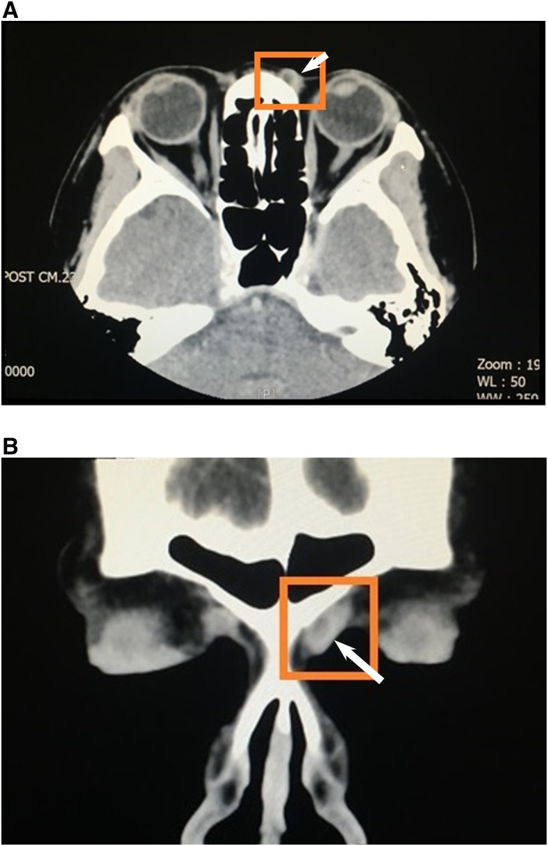
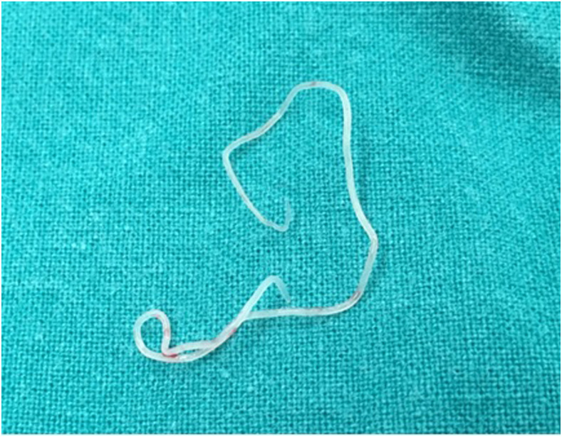
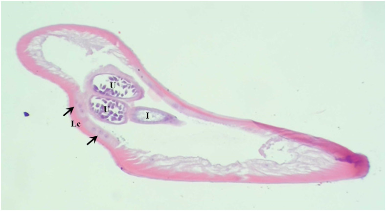
Brugia malayi is a lymphatic nematode that accounts for approximately 10% of lymphatic filariasis cases worldwide. It is endemic in several countries in South and Southeast Asia. In Thailand, B. malayi is endemic in the southern region. The extralymphatic presentation of B. malayi is rare. Here, we report the case of a woman residing in the central region of Thailand who presented with an erythematous periorbital nodule at the left medial canthal area caused by lymphatic filaria. A viable sexually mature filarial adult was removed from the lesion. The nematode species was identified as B. malayi by histology staining and DNA sequencing of the partial mitochondrial 12S ribosomal RNA (rRNA) gene. As far as we know, this is the first case report of B. malayi presenting with a periorbital nodule that has occurred in a disease non-endemic area of Thailand with possibly a zoonotic origin.
Figures
Similar articles
-
Ocular Brugia pahangi Filariasis Complicated by Severe Macular Damage in Thailand: Case Report and Literature Review.Am J Trop Med Hyg. 2024 Apr 30;110(6):1158-1164. doi: 10.4269/ajtmh.24-0047. Print 2024 Jun 5. Am J Trop Med Hyg. 2024. PMID: 38688273 Free PMC article. Review.
-
Detection of filarial parasites in domestic cats by PCR-RFLP of ITS1.Vet Parasitol. 2006 Sep 10;140(3-4):366-72. doi: 10.1016/j.vetpar.2006.04.003. Epub 2006 May 19. Vet Parasitol. 2006. PMID: 16713099
-
A rare case of ocular filariasis caused by Brugia malayi.Parasitol Int. 2022 Oct;90:102606. doi: 10.1016/j.parint.2022.102606. Epub 2022 Jun 2. Parasitol Int. 2022. PMID: 35659633
-
Molecular cloning, purification and characterisation of myosin of human lymphatic filarial parasite Brugia malayi.Parasitol Res. 2008 Feb;102(3):481-9. doi: 10.1007/s00436-007-0786-2. Epub 2007 Dec 7. Parasitol Res. 2008. PMID: 18064491
-
DNA-based diagnosis of lymphatic filariasis.Southeast Asian J Trop Med Public Health. 2009 Sep;40(5):904-13. Southeast Asian J Trop Med Public Health. 2009. PMID: 19842372 Review.
Cited by
-
Current Status of the Diagnosis of Brugia spp. Infections.Pathogens. 2024 Aug 23;13(9):714. doi: 10.3390/pathogens13090714. Pathogens. 2024. PMID: 39338906 Free PMC article. Review.
-
Perspectives of vector management in the control and elimination of vector-borne zoonoses.Front Microbiol. 2023 Mar 21;14:1135977. doi: 10.3389/fmicb.2023.1135977. eCollection 2023. Front Microbiol. 2023. PMID: 37025644 Free PMC article. Review.
-
The first case report of subcutaneous dirofilariasis caused by Dirofilaria repens in Thailand.Trop Parasitol. 2021 Jul-Dec;11(2):125-127. doi: 10.4103/tp.TP_113_20. Epub 2021 Oct 20. Trop Parasitol. 2021. PMID: 34765535 Free PMC article.
-
Integrated Histological and Molecular Analysis of Filarial Species and Associated Wolbachia Endosymbionts in Human Filariasis Cases Presenting Atypically in Thailand.Am J Trop Med Hyg. 2024 Aug 6;111(4):829-840. doi: 10.4269/ajtmh.24-0147. Print 2024 Oct 2. Am J Trop Med Hyg. 2024. PMID: 39106844
-
Ocular Brugia pahangi Filariasis Complicated by Severe Macular Damage in Thailand: Case Report and Literature Review.Am J Trop Med Hyg. 2024 Apr 30;110(6):1158-1164. doi: 10.4269/ajtmh.24-0047. Print 2024 Jun 5. Am J Trop Med Hyg. 2024. PMID: 38688273 Free PMC article. Review.
References
-
- CDC , 2019. CDC - Lymphatic Filariasis - General Information - Vectors of Lypmhatic Filaraisis. Available at: https://www.cdc.gov/parasites/lymphaticfilariasis/gen_info/vectors.html. Accessed October 11, 2019.
-
- Zielke E, Hinz E, 1993. Lymphatic filariasis in Thailand. A review on distribution and transmission. Trop Parasitol 15: 141–148.
-
- World Health Organization , 2017. Elimination of Lymphatic Filariasis as a Public Health Problem in Thailand. Available at: http://www.searo.who.int/thailand/news/elimination-of-lymphatic-filarias.... Accessed August 2, 2018.
-
- Edeson JFB, Wilson T, 1964. The epidemiology of filariasis due to Wuchereria bancrofti and Brugia malayi. Annu Rev Entomol 9: 245–268.
Publication types
MeSH terms
Substances
LinkOut - more resources
Full Text Sources
Research Materials
