Case Reports
. 2020 Dec;103(6):2318-2322.
doi: 10.4269/ajtmh.20-0839. Epub 2020 Sep 17.

Case Report: Neurocysticercosis Acquired in Australia

Affiliations
Case Reports

Case Report: Neurocysticercosis Acquired in Australia

Daniel Forster et al. Am J Trop Med Hyg. 2020 Dec.

Abstract

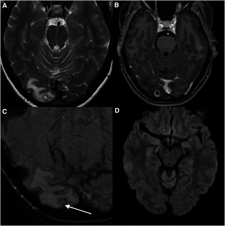
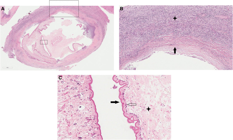
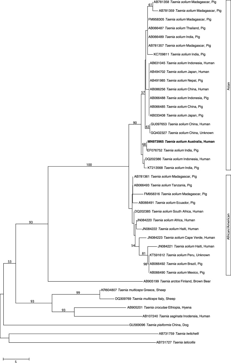
Neurocysticercosis (NCC) is a disease caused by infection of the central nervous system with the larval stage of the tapeworm Taenia solium. This disease is endemic in many parts of the world, including Africa, Asia, and Latin America, where animal husbandry practices are common such that pigs reared for human consumption ingest feces from humans infected with T. solium. Neurocysticercosis is rarely acquired in economically affluent regions, including North America, Central Europe, Japan, and Australasia, and in countries where pork consumption is discouraged by religious or social practices. In these countries, NCC is usually diagnosed in immigrants or returning travelers who have spent time in endemic regions. Here, we report a case of NCC in a 25-year-old woman presenting with worsening visual symptoms in association with headache, diagnosed previously as a migraine with visual aura. This person had always lived in Australia and had never traveled overseas to a country endemic for T. solium. The unusual features of the clinical presentation and epidemiology are highlighted to raise physicians' awareness that attention needs to be paid to the risk of autochthonous infection occurring in non-endemic countries.


Figures

Figure 1.
Figure 1.
Magnetic resonance imaging (MRI) for the detection and localization of the cystic lesion. (A) T2, (B) T1-weighted contrast enhanced, (C) susceptibility-weighted, and (D) diffusion-weighted images from an MRI brain show a cystic mass (8 mm), located peripherally in the right occipital lobe with surrounding vasogenic edema. The cyst had an enhancing T2 hypointense wall. Best appreciated on the T2 and susceptibility-weighted image sequences is a punctate low-signal focus within the cyst (arrow). The lack of diffusion restriction of the fluid contents is not consistent with a pyogenic abscess. The morphology was typical of neurocysticercosis in the colloidal vesicular phase, and the internal punctate focus represents the parasite scolex.
Figure 2.
Figure 2.
Histopathology of the excised cystic lesion. The cyst and adjacent brain tissue stained with hematoxylin and eosin. (A) Cysticercus (×40 magnification) with surrounding fibrous capsule and inflamed brain parenchyma. (B) Fibrous capsule (black arrow; ×100 magnification) and adjacent white matter inflammatory cell infiltrate (star) consisting of lymphocytes, plasma cells, histiocytes, and eosinophils. (C) Cyst wall tegument (×400) demonstrating outer microvilli (black arrow), cellular layer (transparent arrow), and inner reticular layer with excretory canaliculi (star). This figure appears in color at www.ajtmh.org.
Figure 3.
Figure 3.
Phylogenetic relationship of sequences in the mitochondrial gene cytochrome c oxidase subunit 1 (cox-1; 446 bp). The sequence derived from DNA from the cranial cyst of the present patient from Australia (bold-type) and other reference sequences from selected Taenia taxa were subjected to analysis using the neighbor-joining distance tree building method. Individual GenBank accession numbers precede species names. Bootstrap values (based on 2,000 iterations) are located next to supported branches. Taenia laticollis (from lynx) was selected as the outgroup. The scale bar indicates the number of nucleotide substitutions per site.

References

    1. Bowles J, Blair D, McManus DP, 1992. Genetic variants within the genus Echinococcus identified by mitochondrial DNA sequencing. Mol Biochem Parasitol 54: 165–173. - PubMed
    1. Bowles J, McManus DP, 1994. Genetic characterization of the Asian Taenia, a newly described taeniid cestode of humans. Am J Trop Med Hyg 50: 33–44. - PubMed
    1. Kumar S, Stecher G, Li M, Knyaz C, Tamura K, 2018. MEGA X: molecular evolutionary genetics analysis across computing platforms. Mol Biol Evol 35: 1547–1549. - PMC - PubMed
    1. Hughes AJ, Biggs BA, 2002. Parasitic worms of the central nervous system: an Australian perspective. Intern Med J 32: 541–553. - PubMed
    1. Holmes NE, Iles LE, Danks AR, Korman TM, 2010. Neurocysticercosis causing sudden death. Am J Forensic Med Pathol 31: 117–119. - PubMed

Publication types