. 2020 Oct;19(10):e13240.
doi: 10.1111/acel.13240. Epub 2020 Sep 22.

CXCL5-CXCR2 signaling is a senescence-associated secretory phenotype in preimplantation embryos

Affiliations

CXCL5-CXCR2 signaling is a senescence-associated secretory phenotype in preimplantation embryos

Yuta Kawagoe et al. Aging Cell. 2020 Oct.

Abstract

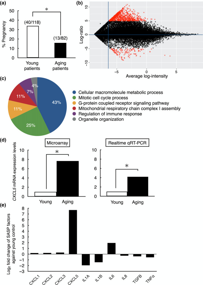
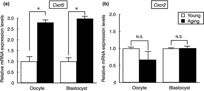
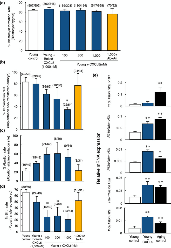
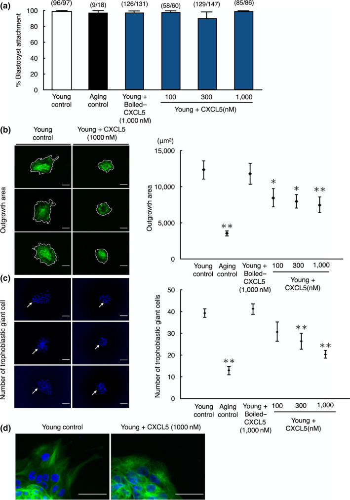
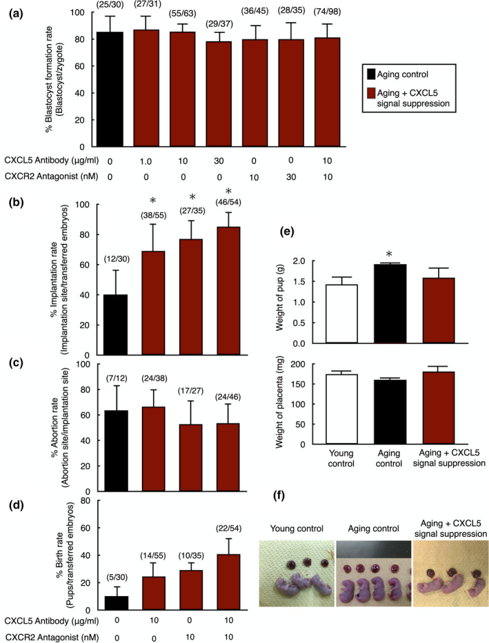
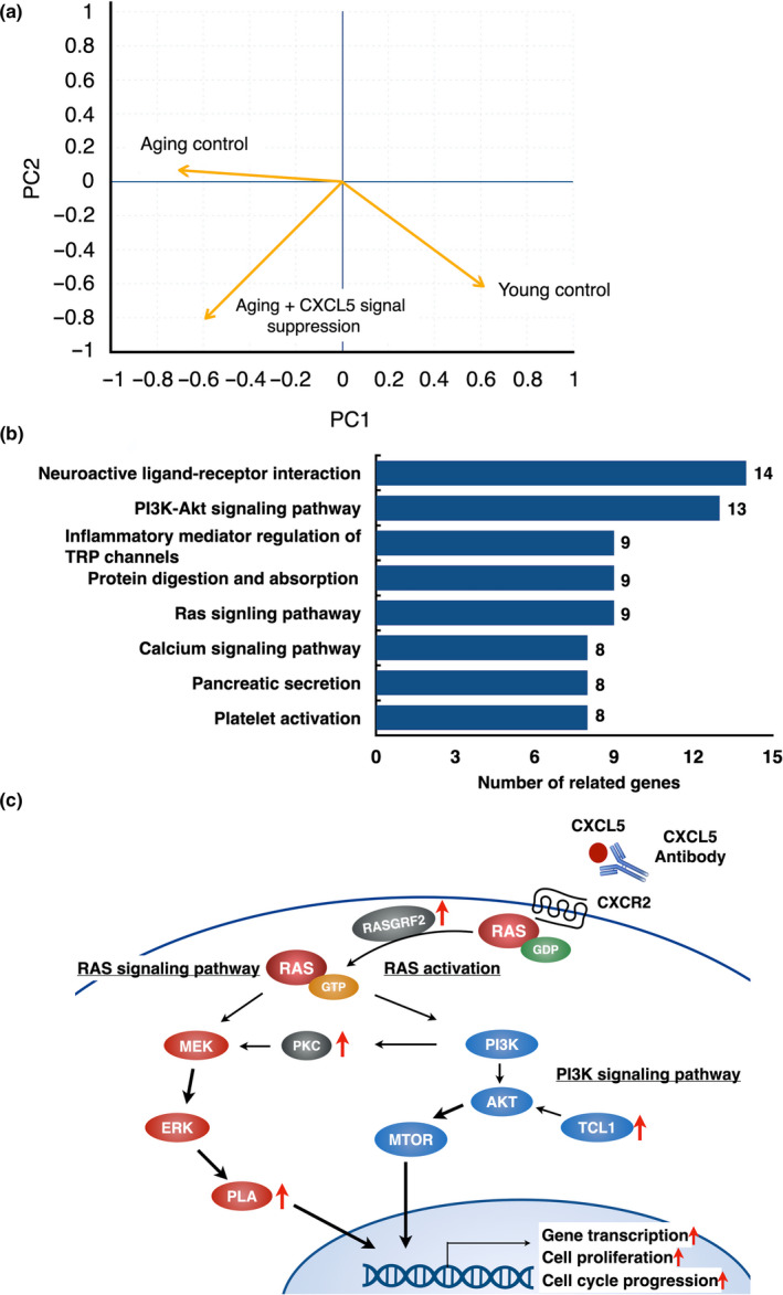
Pregnancy rate of women decreases with age due to declining quality of oocytes and embryos. However, there is no established method to improve pregnancy rate in aging women. In this study, we identified a senescence-associated secretory phenotype (SASP) factor partially responsible for the decline in embryo implantation potential. Based on microarray analysis using young and aging human embryos at the same morphological grade, 702 genes showed >fivefold increases in aging human blastocysts. Among these genes, C-X-C motif chemokine 5 (CXCL5) showed 7.7-fold increases in aging human blastocysts. However, no-age-dependent changes in expression of the CXCR2, the cognate receptor for CXCL5, were found. In aging mice, Cxcl5 transcript levels were also increased in oocytes and embryos. Treatment of young mouse embryos with CXCL5 decreased implantation rates, together with increased expression of aging markers (P53, P21, Pai-1, and Il-6). Moreover, CXCL5 treatment suppressed trophoblast outgrowth in young mouse blastocysts. Conversely, suppression of CXCL5-CXCR2 signaling in aging mouse embryos using neutralizing antibodies and a receptor antagonist improved the implantation rate, leading to increases in pregnancy and delivery of normal pups. The gene expression pattern of these embryos was comparable to that in young mouse embryos showing enriched cell proliferation-related pathways. In conclusion, we identified CXCL5 as a SASP factor in human and mouse embryos and suppression of CXCL5-CXCR2 signaling during embryo culture improved pregnancy success in aging mice. Future analysis on CXCL5-CXCR2 signaling suppression in human embryos could be the basis to improve embryo development and pregnancy outcome in middle-aged infertile patients.

Keywords: CXCL5; CXCR2; SASP; aging; infertility; preimplantation embryo.


Conflict of interest statement

The authors confirm that they have no conflict of interest.

Figures

FIGURE 1
FIGURE 1
Blastocyst gene expression analysis between young and aging patients to identify SASP factors derived from human embryos. (a) Pregnancy rate of young (25–37 years, n = 118) and aging (38–44 years, n = 82) patients after high‐quality blastocyst (above 3BB by the Gardner criteria) transfer to normal thickness of endometrium (>7 mm). *p < 0.01. (b) A scatter plot of all 23,210 transcripts expressed in human blastocysts analyzed using Agilent Whole human V2 genome Oligo microarray 4 × 44 K. Gene expression in two age groups (young; 25–37 years n = 5, aging; 38–44 years n = 5) was compared. Total 820 genes were identified as differentially expressed genes (red marks, fold change >5, p < 0.01); 702 genes were highly expressed in aging blastocysts and 118 genes highly expressed in young blastocysts. (c) GO analysis of differentially expressed genes. Six GO terms based on biological process were significantly over‐represented in aging human blastocysts. (d) Validation of microarray data on CXCL5 expression by using quantitative real‐time RT‐PCR. *p < 0.01. Data of quantitative real‐time RT‐PCR were shown as mean ± SE. n = 3 per group. (e) Expression levels of key SASP factors in human aging blastocysts. Data were presented as fold changes against human young embryos
FIGURE 2
FIGURE 2
Expression of CXCL5 in oocytes and embryos of young and aging mice. (a) Transcript levels for Cxcl5 and (b) Cxcr2, cognate receptor of Cxcl5, in oocytes and blastocysts of young and aging mice. Cxcl5 and Cxcr2 mRNA levels were measured by using real‐time quantitative RT‐PCR. The values of all mRNAs were normalized based on those for Histon H2a in the same samples. *p < 0.01, N.S.: not significant difference. All results were shown as mean ± SE, n = 3 per group
FIGURE 3
FIGURE 3
Negative impacts of CXCL5 treatment on early embryo implantation and live birth from young mice. (a) Blastocyst formation rate of young embryos treated with CXCL5 peptide. Oocyte from young mice was fertilized in vitro and allowed to develop into blastocysts with or without CXCL5 peptide treatment. The number of embryos developed to blastocyst vs. total number of embryos are listed on top of each column. Data were shown as mean ± SE n = 10–15 per group. (b‐d) Implantation (b), abortion (c), and birth rates (d) of young embryos treated with CXCL5 peptide. Cultured blastocysts were transferred to the uterus of recipient mice. On day 19.5 of pregnancy, all recipients were sacrificed to the number of pups, the implantation sites and the aborted fetuses were counted. The numbers listed on top of each column indicates the number of implantation site/total number of blastocysts transferred, the number of abortion site/the number of implantation sites and the number of pups/total number of blastocysts transferred, respectively. Data were shown as mean ± SE n = 10–15/group. *p < 0.01, vs. young control. In all experiments, boiled‐CXCL5 at 1,000 nM was served as negative control (Boiled‐CXCL5), whereas anti‐CXCL5 neutralizing antibody and CXCR2 selective antagonist were used to block CXCL5 signaling (Antibody + Antagonist). Ab, Antibody; An, Antagonist. (e) Expression levels of aging markers (P16, P21, P53, Pai1, and Il6) in young embryos with or without treatment with CXCL5 and in aging embryos. Levels of aging markers were measured by real‐time quantitative RT‐PCR. The values were normalized based on those for Histon H2a in the same sample. Data were shown as mean ± SE. N = 5–7 per group. *p < 0.05 vs. young control. **p < 0.01 vs. young control
FIGURE 4
FIGURE 4
Decreases in blastocyst outgrowth without affecting blastocyst attachment in young mouse embryos by treatment with CXCL5. (a) Blastocyst attachment rate, (b) outgrowth area, and (c) number of trophoblastic giant cells of young embryos treated with CXCL5 peptide. (a) The ability of embryo attachment to culture dishes was evaluated by culturing blastocysts for an additional three days. The numbers listed on top of each column indicated the number of attached embryo/total number of blastocysts cultured. (b) Outgrowth was identified by existence of trophoblastic giant cells stained with α‐Tubulin on the culture dishes. The enclosed areas on left panels indicated outgrowth area for each group. (c) For counting trophoblastic giant cells, left panels were representative images of nuclear stained embryos. Arrows indicated trophoblastic giant cells. (d) Representative magnified images of trophoblastic giant cells at peripheral area of outgrowth for each group. Data were shown as mean ± SE. N = 5–8 per group. *p < 0.05 vs. young control. **p < 0.01 vs. young control. Boiled‐CXCL5 at 1,000 nM was served as negative control (Boiled‐CXCL5), whereas blastocysts from aging mice (Aging) were used for positive control. Bars: 100 µm
FIGURE 5
FIGURE 5
Increases in pregnancy success of aging mouse embryos with suppression of CXCL5‐CXCR2 signaling before embryo transfer. (a) Blastocyst formation rate of aging embryos with suppression of CXCL5‐CXCR2 signaling. In vitro fertilized zygotes from aging mice were allowed to develop into blastocysts without or with anti‐CXCL5 neutralizing antibody and/or CXCR2 antagonist. The number of embryos developed to blastocyst vs. total number of embryos are listed on top of each column. Data were shown as mean ± SE. N = 10–15 per group. (b–d) Implantation (b), abortion (c), and birth rates (d) of aging embryos treated with anti‐CXCL5 neutralizing antibody and/or CXCR2 antagonist. Cultured blastocysts were transferred to the uterus of recipient mice. On day 19.5 of pregnancy, the number of pups, the implantation sites, and the aborted fetuses were counted by sacrificing animals. The numbers listed on top of each column indicated the number of implantation site/total number of blastocysts transferred, the number of abortion site/the number of implantation site, and the number of pups/total number of blastocysts transferred, respectively. (e) Effects of CXCL5‐CXCR2 signaling suppression on pups and placentas derived from aging embryos. The weight of pups and placentas were measured on day 19.5 of pregnancy by sacrificing animals. (f) Representative images of pups and placentas in each group. Data were shown as mean ± SE. N = 10‐20 per group. *p < 0.01, vs. young control. Data were shown as mean ± SE. N = 10–15 per group. *p < 0.01, vs. aging control without anti‐CXCL5 neutralizing antibody and/or CXCR2 antagonist treatment
FIGURE 6
FIGURE 6
Analysis of differentially expressed genes between aging mouse blastocysts with and without CXCL5 signaling suppression. (a) Similarity of gene expression pattern of embryos between young and aging mouse blastocysts with or without CXCL5 signaling suppression. Using blastocysts samples, principal component (PC) analysis using data of microarray analysis was performed. (b) KEGG (Kyoto Encyclopedia of Genes and Genomes) pathway analysis on differentially expressed genes in aging embryos with or without CXCL5 signal suppression using data of microarray analysis. The graph showed the enriched pathways. Numbers listed on left of each column indicate the number of related genes. (c) Schematic diagram of enriched signaling pathway following suppression of CXCL5 signaling. PI3 K‐AKT and RAS signaling pathway were affected by CXCL5 signaling suppression. The genes located besides the red arrows are enriched and significantly up‐regulated in CXCL5‐suppressed aging blastocysts

Similar articles

Cited by

References

    1. Acosta, J. C. , Banito, A. , Wuestefeld, T. , Georgilis, A. , Janich, P. , Morton, J. P. , … Gil, J. (2013). A complex secretory program orchestrated by the inflammasome controls paracrine senescence. Nature Cell Biology, 15(8), 978–990. 10.1038/ncb2784 - DOI - PMC - PubMed
    1. Acosta, J. C. , O'Loghlen, A. , Banito, A. , Guijarro, M. V. , Augert, A. , Raguz, S. , … Gil, J. (2008). Chemokine signaling via the CXCR2 receptor reinforces senescence. Cell, 133(6), 1006–1018. 10.1016/j.cell.2008.03.038 - DOI - PubMed
    1. Arenberg, D. A. , Keane, M. P. , DiGiovine, B. , Kunkel, S. L. , Morris, S. B. , Xue, Y. Y. , … Strieter, R. M. (1998). Epithelial‐neutrophil activating peptide (ENA‐78) is an important angiogenic factor in non‐small cell lung cancer. Journal of Clinical Investigation, 102(3), 465–472. 10.1172/jci3145 - DOI - PMC - PubMed
    1. Balamayooran, G. , Batra, S. , Cai, S. , Mei, J. , Worthen, G. S. , Penn, A. L. , & Jeyaseelan, S. (2012). Role of CXCL5 in leukocyte recruitment to the lungs during secondhand smoke exposure. American Journal of Respiratory Cell and Molecular Biology, 47(1), 104–111. 10.1165/rcmb.2011-0260OC - DOI - PMC - PubMed
    1. Banito, A. , & Lowe, S. W. (2013). A new development in senescence. Cell, 155(5), 977–978. 10.1016/j.cell.2013.10.050 - DOI - PMC - PubMed

Publication types