. 2020 Dec;7(6):3772-3781.
doi: 10.1002/ehf2.12958. Epub 2020 Sep 22.

Fatal lymphocytic cardiac damage in coronavirus disease 2019 (COVID-19): autopsy reveals a ferroptosis signature

Affiliations

Fatal lymphocytic cardiac damage in coronavirus disease 2019 (COVID-19): autopsy reveals a ferroptosis signature

Werner Jacobs et al. ESC Heart Fail. 2020 Dec.

Abstract

Aims: Cardiovascular complications, including myocarditis, are observed in coronavirus disease 2019 (COVID-19). Major cardiac involvement is a potentially lethal feature in severe cases. We sought to describe the underlying pathophysiological mechanism in COVID-19 lethal cardiogenic shock.

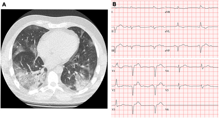
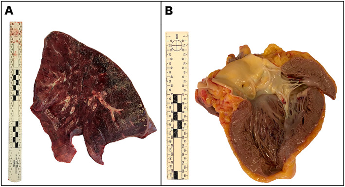
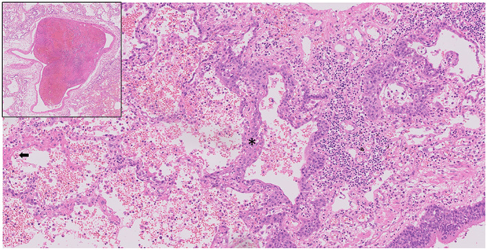
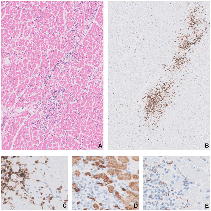
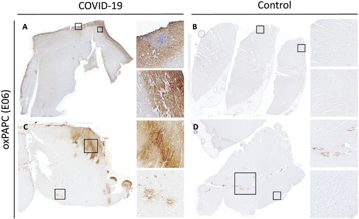
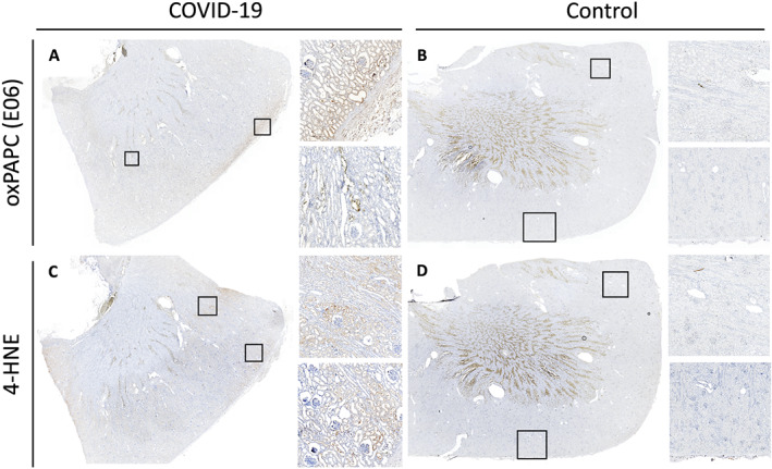
Methods and results: We report on a 48-year-old male COVID-19 patient with cardiogenic shock; despite extracorporeal life support, dialysis, and massive pharmacological support, this rescue therapy was not successful. Severe acute respiratory syndrome coronavirus 2 RNA was detected at autopsy in the lungs and myocardium. Histopathological examination revealed diffuse alveolar damage, proliferation of type II pneumocytes, lymphocytes in the lung interstitium, and pulmonary microemboli. Moreover, patchy muscular, sometimes perivascular, interstitial mononuclear inflammatory infiltrates, dominated by lymphocytes, were seen in the cardiac tissue. The lymphocytes 'interlocked' the myocytes, resulting in myocyte degeneration and necrosis. Predominantly, T-cell lymphocytes with a CD4:CD8 ratio of 1.7 infiltrated the interstitial myocardium, reflecting true myocarditis. The myocardial tissue was examined for markers of ferroptosis, an iron-catalysed form of regulated cell death that occurs through excessive peroxidation of polyunsaturated fatty acids. Immunohistochemical staining with E06, a monoclonal antibody binding to oxidized phosphatidylcholine (reflecting lipid peroxidation during ferroptosis), was positive in morphologically degenerating and necrotic cardiomyocytes adjacent to the infiltrate of lymphocytes, near arteries, in the epicardium and myocardium. A similar ferroptosis signature was present in the myocardium of a COVID-19 subject without myocarditis. In a case of sudden death due to viral myocarditis of unknown aetiology, however, immunohistochemical staining with E06 was negative. The renal proximal tubuli stained positively for E06 and also hydroxynonenal (4-HNE), a reactive breakdown product of the lipid peroxides that execute ferroptosis. In the case of myocarditis of other aetiology, the renal tissue displayed no positivity for E06 or 4-HNE.

Conclusions: The findings in this case are unique as this is the first report on accumulated oxidized phospholipids (or their breakdown products) in myocardial and renal tissue in COVID-19. This highlights ferroptosis, proposed to detrimentally contribute to some forms of ischaemia-reperfusion injury, as a detrimental factor in COVID-19 cardiac damage and multiple organ failure.

Keywords: Autopsy; COVID-19; Ferroptosis; Lymphocytic myocarditis; Renal failure; SARS-CoV-2-infection.


Conflict of interest statement

The findings and conclusions in this report are those of the authors. The authors have nothing to disclose.

Figures

FIGURE 1
FIGURE 1
(A) Chest CT. The coronal image shows multiple patchy ground‐glass opacifications in all lung fields (right more than left) (Illness Day 7, Hospital Day 1). (B) Electrocardiogram on Day 5 after admission showing a widened QRS in the absence of hypokalaemia (Illness Day 11, Hospital Day 5).
FIGURE 2
FIGURE 2
Macroscopy at autopsy. (A) The lungs were purplish, edematous, and had a mottled aspect. (B) View on the left ventricle with some petechiae in the endocardium.
FIGURE 3
FIGURE 3
Lung tissue with diffuse alveolar damage, with hyaline formation (arrow), marked type 2 pneumocyte hyperplasia (asterisk) with prominent nucleoli and multinucleation and interstitial mononuclear inflammatory infiltrates (predominantly with lymphocytes) (original magnification ×100). Insert: microembolus.
FIGURE 4
FIGURE 4
Histochemical and immunohistochemical examination of a heart tissue sample (several sections of the heart performed: septum n = 2, anterior wall n = 2, posterior wall n = 2, right ventricle n = 1). Lymphocytes infiltrating the cardiomyocytes (A–C), resulting in myocyte degeneration and necrosis (D). Most of the lymphocytes are CD3‐positive (B) and CD4‐positive (C) T‐cells. The CD4‐positive cells progressively infiltrate the cardiomyocytes (C) demonstrated by the progressive disappearance of the desmin‐positive myofibrils (brown in panel D) and are accompanied with only a few CD68‐positive macrophages (E). Staining: HE (A), CD3 (B), CD4 (C), desmin (D), and CD68 (E). Original magnification: ×50 (A, B) and ×200 (C, D).
FIGURE 5
FIGURE 5
Immunohistochemical examination of heart tissue for a ferroptosis marker. E06 immunoreactivity (protocol adapted from Haider et al. 9 ) was detected in the area of severe SARS‐CoV‐2 myocarditis, near arteries and throughout the myocardium in a gradient‐like manner (A). In a case of a 16‐year‐old man who suddenly died of non‐COVID‐19 myocarditis due to other aetiology, no signal was detected on staining with E06 (B). The myocardium of a 33‐year‐old male COVID‐19 patient that succumbed to severe acute respiratory distress syndrome and multiple organ failure, but without myocarditis, showed foci of E06 positivity in all areas of the ventricular wall and perivascular regions (C). The myocardium of another control subject without myocarditis, a 48‐year‐old man who was hospitalized after trauma, showed only some E06 positivity in perivascular connective tissue (D). Control stains using only secondary antibodies are available in the Supporting Information.
FIGURE 6
FIGURE 6
Immunohistochemical examination of renal tissue for potential ferroptosis markers. Renal tissue from the COVID‐19 patient with myocarditis and multiple organ dysfunction syndrome showed morphological signs of acute tubular necrosis, intratubular oxalate crystals, as well as E06 positivity in proximal tubuli (A). The latter also stained positively for the presence of 4‐HNE, one of the breakdown product of lipid peroxides (protocol adapted from Feng et al. 11 ) (C). By comparison, in the case of sudden death due to myocarditis of other aetiology, immunohistochemical staining with E06 (B) and anti‐4‐HNE antibody (D) in the renal tissue showed no presence of these ferroptosis markers (non‐specific staining in the corticomedullary junction is also present on control stains). Control stains using only secondary antibodies are enclosed in the Supporting Information.

References

    1. Zhu N, Zhang D, Wang W, Zhu N, Zhang D, Wang W, Li X, Yang B, Song J, Zhao X, Huang B, Shi W, Lu R, Niu P. A novel coronavirus from patients with pneumonia in China, 2019. N Engl J Med 2020; 382: 727–733. - PMC - PubMed
    1. Guo T, Fan Y, Chen M, Wu X, Zhang L, He T, Wang H, Wan J, Wang X, Lu Z. Cardiovascular implications of fatal outcomes of patients with coronavirus disease 2019 (COVID‐19). JAMA Cardiol 2020; 5: 811. - PMC - PubMed
    1. Veronese G, Ammirati E, Cipriani M, Frigerio M. Fulminant myocarditis: characteristics, treatment, and outcomes. Anatol J Cardiol 2018; 19: 279–286. - PMC - PubMed
    1. Ackermann M, Verleden SE, Kuehnel M, Haverich A, Welte T, Laenger F, Vanstapel A, Werlein C, Stark H, Tzankov A, Li WW, Li VW, Mentzer SJ, Jonigk D. Pulmonary vascular endothelialitis, thrombosis, and angiogenesis in COVID‐19. N Engl J Med 2020; 383: 120–128. - PMC - PubMed
    1. Corman VM, Landt O, Kaiser M, Molenkamp R, Meijer A, Chu DKW, Bleicker T, Brünink S, Schneider J, Schmidt ML, Mulders DGJC, Haagmans BL, van der Veer B, van den Brink S, Wijsman L, Goderski G, Romette JL, Ellis J, Zambon M, Peiris M, Goossens H, Reusken C, Koopmans MPG, Drosten C. Detection of 2019 novel coronavirus (2019‐nCoV) by real‐time RT‐PCR. Euro Surveill 2020; 25: 2000045. - PMC - PubMed