. 2020 Oct 9;48(18):10567-10575.
doi: 10.1093/nar/gkaa738.

Duplex formation in a G-quadruplex bulge

Affiliations

Duplex formation in a G-quadruplex bulge

Thi Quynh Ngoc Nguyen et al. Nucleic Acids Res. .

Abstract

Beyond the consensus definition of G-quadruplex-forming motifs with tracts of continuous guanines, G-quadruplexes harboring bulges in the G-tetrad core are prevalent in the human genome. Here, we study the incorporation of a duplex hairpin within a bulge of a G-quadruplex. The NMR solution structure of a G-quadruplex containing a duplex bulge was resolved, revealing the structural details of the junction between the duplex bulge and the G-quadruplex. Unexpectedly, instead of an orthogonal connection the duplex stem was observed to stack below the G-quadruplex forming a unique quadruplex-duplex junction. Breaking up of the immediate base pair step at the junction, coupled with a narrowing of the duplex groove within the context of the bulge, led to a progressive transition between the quadruplex and duplex segments. This study revealed that a duplex bulge can be formed at various positions of a G-quadruplex scaffold. In contrast to a non-structured bulge, the stability of a G-quadruplex slightly increases with an increase in the duplex bulge size. A G-quadruplex structure containing a duplex bulge of up to 33 nt in size was shown to form, which was much larger than the previously reported 7-nt bulge. With G-quadruplexes containing duplex bulges representing new structural motifs with potential biological significance, our findings would broaden the definition of potential G-quadruplex-forming sequences.


Figures

Figure 1.
Figure 1.
Schematics of a G-quadruplex containing a single-nucleotide bulge (left) and a duplex bulge (right).
Figure 2.
Figure 2.
1D imino proton NMR spectra of (A) B4-dx2, (B) B4-T and (C) dx2. Imino protons of the G-tetrad core are indicated by filled circles, whereas imino protons of the duplex are indicated by open squares. (D) CD spectra of B4-T (green), dx2 (blue) and B4-dx2 (red).
Figure 3.
Figure 3.
NMR structural characterization of B4-dx2. (A) NOESY spectrum (mixing time, 200 ms) showing cross-peaks for the identification of three G-tetrads and five Watson-Crick base pairs. Signature NOE cross-peaks for the Watson-Crick base pairs and G-tetrads are framed and labeled with the residue numbers of the imino and H8 protons at the first and second positions, respectively. (B) NOE between the thymine imino proton and the adenine H2 proton in an A•T base pair. (C) NOEs between the guanine imino proton and the cytosine amino protons in a G•C base pair. (D) NOEs between H8 and imino protons within a G-tetrad. (E) Schematic diagram of B4-dx2.
Figure 4.
Figure 4.
NMR solution structure of B4-dx2. (A) Ten superimposed lowest-energy structures. (B) Ribbon view of a representative structure. Anti guanines in the G-tetrads are colored in cyan; syn guanine in the G-tetrads, green; bases in the duplex stem, magenta; backbone and sugar, gray; O4′ atoms, red; phosphorus atoms, yellow.
Figure 5.
Figure 5.
Structural features of B4-dx2 at the quadruplex–duplex junction. (A) Stacking between T19 (magenta) and G20 (green). (B) Backbone strand separation at the junction for the duplex stem and the G-tetrad core (C) Stretching of backbone between the G4 and A5 residues.
Figure 6.
Figure 6.
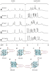
1D imino proton NMR spectra and schematics of G-quadruplexes containing a duplex bulge of different sizes: (A, A’) B4-dx2 (15 nt), (B, B’) B4-dx3 (25 nt) and (C, C’) B4-dx4 (33 nt).
Figure 7.
Figure 7.
1D imino proton NMR spectra and schematics of quadruplex–duplex hybrids with a duplex hairpin inserted at different bulge positions: (A, A’) B3-dx2 with duplex at the bulge position 3, (B, B’) B4-dx2 with duplex at the bulge position 4, (C, C’) B7-dx2 with duplex at the bulge position 7, (D, D’) B8-dx2 with duplex at the bulge position 8, and (E, E’) B12-dx2 with duplex at the bulge position 12.

References

    1. Davis J.T. G-quartets 40 years later: from 5′-GMP to molecular biology and supramolecular chemistry. Angew. Chem. Int. Ed. Engl. 2004; 43:668–698. - PubMed
    1. Burge S., Parkinson G.N., Hazel P., Todd A.K., Neidle S.. Quadruplex DNA: sequence, topology and structure. Nucleic. Acids. Res. 2006; 34:5402–5415. - PMC - PubMed
    1. Patel D.J., Phan A.T., Kuryavyi V.. Human telomere, oncogenic promoter and 5′-UTR G-quadruplexes: diverse higher order DNA and RNA targets for cancer therapeutics. Nucleic. Acids. Res. 2007; 35:7429–7455. - PMC - PubMed
    1. Gellert M., Lipsett M.N., Davies D.R.. Helix formation by guanylic acid. Proc. Natl. Acad. Sci. U.S.A. 1962; 48:2013–2018. - PMC - PubMed
    1. Sen D., Gilbert W.. Formation of parallel four-stranded complexes by guanine-rich motifs in DNA and its implications for meiosis. Nature. 1988; 334:364–366. - PubMed

Publication types