. 2021 Jun;15(3):1589-1621.
doi: 10.1007/s11682-020-00357-9.

MRI-based Parcellation and Morphometry of the Individual Rhesus Monkey Brain: the macaque Harvard-Oxford Atlas (mHOA), a translational system referencing a standardized ontology

Affiliations

MRI-based Parcellation and Morphometry of the Individual Rhesus Monkey Brain: the macaque Harvard-Oxford Atlas (mHOA), a translational system referencing a standardized ontology

R Jarrett Rushmore et al. Brain Imaging Behav. 2021 Jun.

Abstract

Investigations of the rhesus monkey (Macaca mulatta) brain have shed light on the function and organization of the primate brain at a scale and resolution not yet possible in humans. A cornerstone of the linkage between non-human primate and human studies of the brain is magnetic resonance imaging, which allows for an association to be made between the detailed structural and physiological analysis of the non-human primate and that of the human brain. To further this end, we present a novel parcellation method and system for the rhesus monkey brain, referred to as the macaque Harvard-Oxford Atlas (mHOA), which is based on the human Harvard-Oxford Atlas (HOA) and grounded in an ontological and taxonomic framework. Consistent anatomical features were used to delimit and parcellate brain regions in the macaque, which were then categorized according to functional systems. This system of parcellation will be expanded with advances in technology and, like the HOA, will provide a framework upon which the results from other experimental studies (e.g., functional magnetic resonance imaging (fMRI), physiology, connectivity, graph theory) can be interpreted.

Keywords: HOA; MRI; Ontology; cortical parcellation; mHOA; macaque monkey.


Figures

Figure 1.
Figure 1.
The state of neuroanatomical knowledge in the individual monkey brain. A. Tract tracing data have been accumulated to reveal the connections of most areas of the monkey brain. These invasive methodologies reveal the origin, stem and termination of axonal bundles, and also illuminate divisions in cortical and subcortical structure by the use of histological stains applied to adjacent or the same brain sections as those for tract tracing. Importantly, these methods do not apply to the in vivo brain. B. Non-invasive diffusion MRI tractography is a method to infer the presence of fiber tracts based on the diffusion of water molecules within white matter. These methods do not have the capability of revealing origins and terminations, but are capable of illustrating stems of fiber pathways. C. MRI-based parcellations divide brain regions, but, by themselves, do not specify origins, terminations or stems of pathways. The three methods together are needed to define the complete brain circuit diagram of the individual monkey in vivo. Upper left figure in A is copyright 2007 Society for Neuroscience and is used with permission; lower left figure in A is used with permission from Petrides and Pandya (1988). Right hand figure in A, and figure B are used with permission from Schmahmann et al. 2007.
Figure 2.
Figure 2.
The state of neuroanatomical knowledge in the individual human brain. A. Since human brains in vivo are not amenable to the use of invasive tract tracing techniques, direct knowledge of pathways is derived from post-mortem microdissection, and from classical histological studies. These methods do not visualize origins or terminations and are validated only for pathway stems. B. Diffusion MRI tractography methods allow for the inference of pathways, but like the same techniques in the monkey (see Figure 1A), provide validated data only for stems. C. Cortical parcellation schemes, such as that of the Harvard-Oxford Atlas, are specified in the individual brain based on consistent anatomical landmarks. These schemes can form the foundation for connectional data derived from non-invasive methods, but do not provide validation of terminations and origins of pathways. Unlike the monkey, combining these three methods in the human are insufficient to define the complete human brain circuit diagram. Left hand figure in A, and figures in B used with permission from Hau et al., 2012. Central and right-hand figures in A from Dejerine and Dejerine-Klumpe (1895).
Figure 3.
Figure 3.
The combination and sequence of approaches in the monkey (Figure 1) that will validate diffusion MRI tractographic technology in the context of cortical parcellation (upper dashed box) and will complete the non-human primate brain circuit diagram in vivo (left). The same approach in the human (right) will lead to an incomplete brain circuit diagram because of the inability to visualize terminations or origins of pathways. However, the validated and confirmed monkey brain circuit diagram will be able to produce a more complete human brain circuit diagram (lower dashed box) through homological comparisons. The dashed arrow indicates the absence of validated origins and terminations of fiber pathways in the human brain (see, e.g., Rushmore et al. 2020).
Figure 4:
Figure 4:
System of Parcellation. An individual T1 image set (left) was acquired and a cortical ribbon and subcortical structures were divided (middle) through a general segmentation process (green – cortical ribbon, orange, cerebellar cortex, yellow – cerebellar white matter, blue- fourth ventricle, blue-grey – brainstem). The cortical ribbon was further divided into regions according to the system described in this paper (right): dark blue – CGP; dark gray – ITG; brown – VMO; gray – STG; light blue – LPCs; light brown – MPC; light green – LPCi; white- PRL.
Figure 5.
Figure 5.
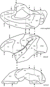
The sulci of the macaque monkey brain from the mid-sagittal (upper), lateral (middle), and ventral (lower) views. The cerebral cortex within the lateral fissure (between the asterisks) is opened to show the constituent regions (to the right of the lateral view). Please note that the arcuate sulcus has a posterior extension (the spur). Abbreviations: arc: arcuate sulcus, cas: callosal sulcus, ccs: calcarine sulcus, cgs: cingulate sulcus, iccs: inferior calcarine sulcus, iocs: inferior occipital sulcus, itps: intraparietal sulcus, crs: limiting sulcus of the insula, lf: lateral fissure, ls: lunate sulcus, ms: marginal sulcus, olfs: olfactory sulcus, ots: occipitotemporal sulcus, pos: parieto-occipital sulcus, prs: principal sulcus, ros: rostral sulcus, rhs: rhinal sulcus, sccs: superior calcarine sulcus, sbps: subparietal sulcus, sts: superior temporal sulcus
Figure 6.
Figure 6.
Parcellation units (PUs) in the mid-sagittal (upper), lateral (middle), and ventral (lower) views. The vertical lines (A-L) represent coronal limiting planes, and non-coronal planes are denoted by lower-case letters (a-c). Areas within the insular cortex are displayed in shading. Abbreviations: CGa:anterior cingulate, CGp:posterior cingulate, COa: central operculum- anterior, COp: central operculum – posterior, F1dl: dorso-lateral superior frontal, F1dm: dorso-medial superior frontal, F2: inferior frontal gyrus, FOC: fronto-orbital cortex, FP: frontal pole, ITG: inferior temporal gyrus, LPCi: inferior portion of lateral parietal cortex, LPCs: superior portion of lateral parietal cortex, MPC: medial parietal cortex, PO: parietal operculum, PoG: postcentral gyrus, PRL: prelunate gyrus, PHG: parahippocampal gyrus, PrG: precentral gyrus, SC: subcallosal area, STG: superior temporal gyrus, STP: supratemporal plane, STRdl: dorsolateral portion of striate cortex, STRm: medial portion of striate cortex, TP: temporal pole, VMO: ventromedial occipital
Figure 7:
Figure 7:
Three-dimensional view of parcellation units in the four cardinal views of the macaque brain. Abbreviations as in Figure 6.
Figure 8.
Figure 8.
Visual representation of relationships among structures (regions) tabulated in Table 4. Parcellation units are boxed to the right of the anatomical structure with which they are associated.
Figure 8.
Figure 8.
Visual representation of relationships among structures (regions) tabulated in Table 4. Parcellation units are boxed to the right of the anatomical structure with which they are associated.

Similar articles

Cited by

References

    1. Aggleton JP, Wright NF, Vann SD, & Saunders RC (2012). Medial temporal lobe projections to the retrosplenial cortex of the macaque monkey. Hippocampus, 22(9), 1883–1900. doi:10.1002/hipo.22024 - DOI - PMC - PubMed
    1. Akbarian S, Grüsser OJ, & Guldin WO (1994). Corticofugal connections between the cerebral cortex and brainstem vestibular nuclei in the macaque monkey. The Journal of Comparative Neurology, 339(3), 421–437. doi:10.1002/cne.903390309 - DOI - PubMed
    1. Allman JM, Tetreault NA, Hakeem AY, Manaye KF, Semendeferi K, Erwin JM, Park S, Goubert V, & Hof PR (2010). The von Economo neurons in frontoinsular and anterior cingulate cortex in great apes and humans. Brain Structure & Function, 214(5–6), 495–517. doi:10.1007/s00429-010-0254-0 - DOI - PubMed
    1. Allman JM, Tetreault NA, Hakeem AY, Manaye KF, Semendeferi K, Erwin JM, et al. (2011). The von Economo neurons in the frontoinsular and anterior cingulate cortex. Annals of the New York Academy of Sciences, 1225, 59–71. doi:10.1111/j.1749-6632.2011.06011.x - DOI - PMC - PubMed
    1. Amaral DG, Insausti R, & Cowan WM (1987). The entorhinal cortex of the monkey: I. Cytoarchitectonic organization. The Journal of Comparative Neurology, 264(3), 326–355. doi:10.1002/cne.902640305 - DOI - PubMed