. 2020 Dec;177(24):5609-5621.
doi: 10.1111/bph.15271. Epub 2020 Oct 21.

Effects of mavacamten on Ca2+ sensitivity of contraction as sarcomere length varied in human myocardium

Affiliations

Effects of mavacamten on Ca2+ sensitivity of contraction as sarcomere length varied in human myocardium

Peter O Awinda et al. Br J Pharmacol. 2020 Dec.

Abstract

Background and purpose: Heart failure can reflect impaired contractile function at the myofilament level. In healthy hearts, myofilaments become more sensitive to Ca2+ as cells are stretched. This represents a fundamental property of the myocardium that contributes to the Frank-Starling response, although the molecular mechanisms underlying the effect remain unclear. Mavacamten, which binds to myosin, is under investigation as a potential therapy for heart disease. We investigated how mavacamten affects the sarcomere-length dependence of Ca2+ -sensitive isometric contraction to determine how mavacamten might modulate the Frank-Starling mechanism.

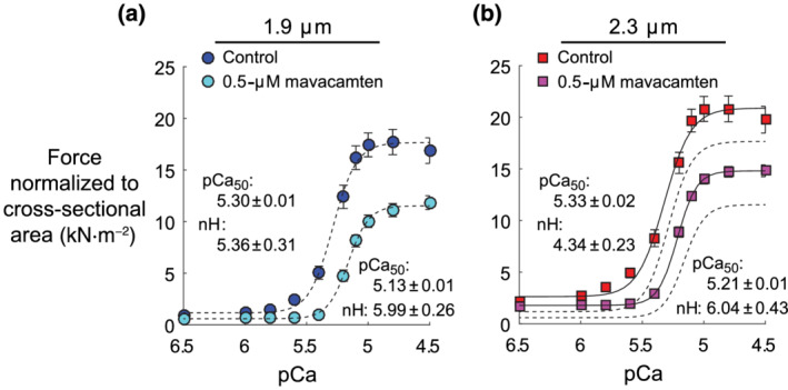
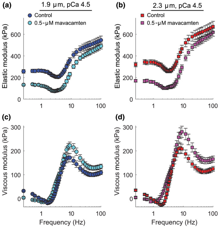
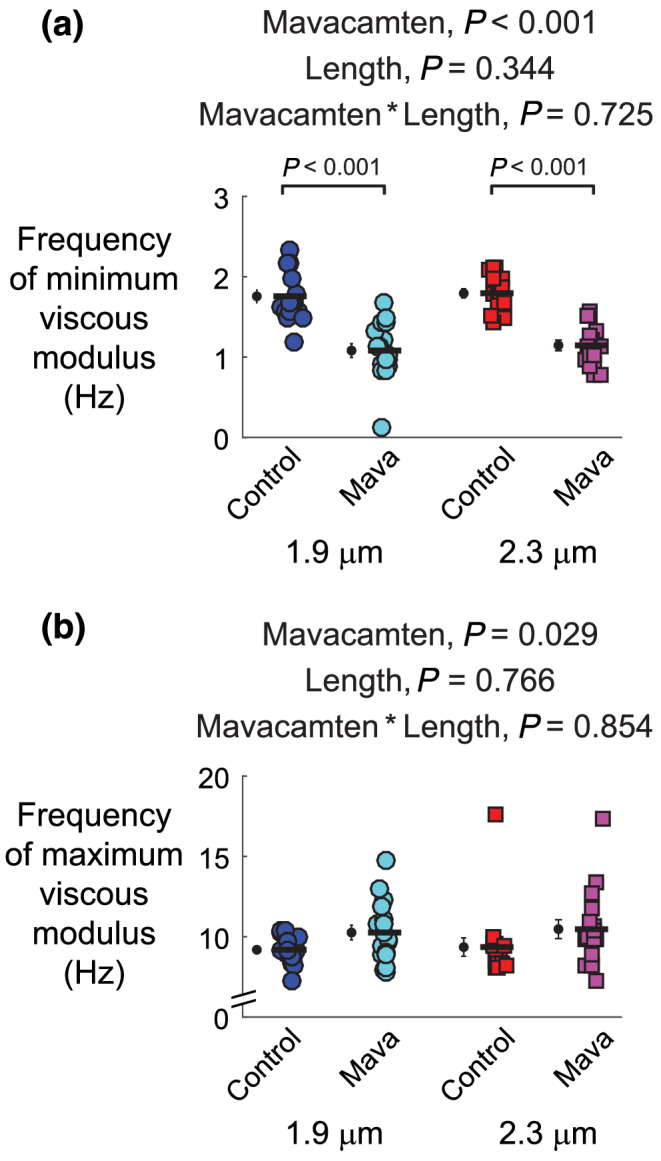
Experimental approach: Multicellular preparations from the left ventricular-free wall of hearts from organ donors were chemically permeabilized and Ca2+ activated in the presence or absence of 0.5-μM mavacamten at 1.9 or 2.3-μm sarcomere length (37°C). Isometric force and frequency-dependent viscoelastic myocardial stiffness measurements were made.

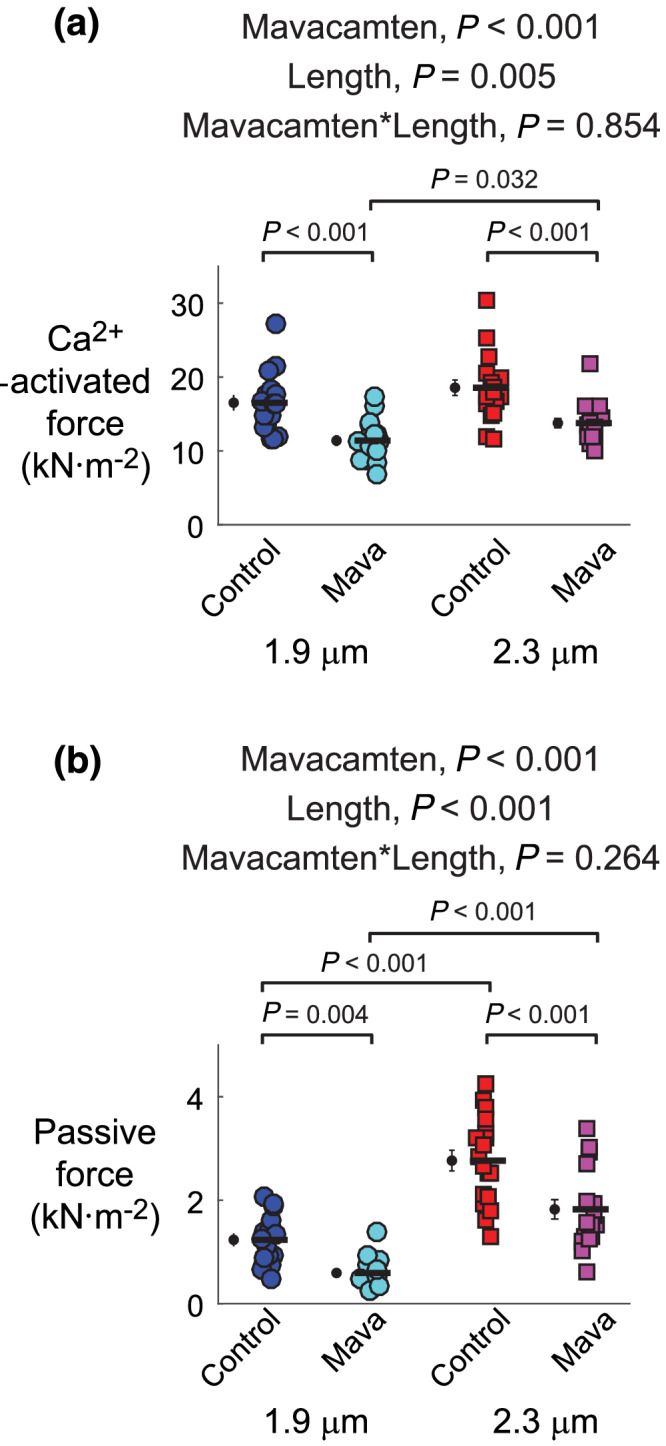
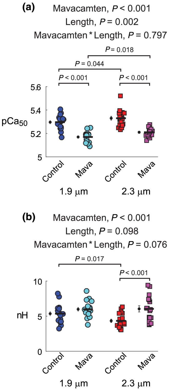
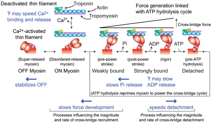
Key results: At both sarcomere lengths, mavacamten reduced maximal force and Ca2+ sensitivity of contraction. In the presence and absence of mavacamten, Ca2+ sensitivity of force increased as sarcomere length increased. This suggests that the length-dependent activation response was maintained in human myocardium, even though mavacamten reduced Ca2+ sensitivity. There were subtle effects of mavacamten reducing force values under relaxed conditions (pCa 8.0), as well as slowing myosin cross-bridge recruitment and speeding cross-bridge detachment under maximally activated conditions (pCa 4.5).

Conclusion and implications: Mavacamten did not eliminate sarcomere length-dependent increases in the Ca2+ sensitivity of contraction in myocardial strips from organ donors at physiological temperature. Drugs that modulate myofilament function may be useful therapies for cardiomyopathies.

Keywords: cardiac muscle mechanics; human myosin; mavacamten; sarcomere length.


Conflict of interest statement

The authors declare no competing financial interests and have nothing to disclose.

Figures

FIGURE 1
FIGURE 1
Schematic introducing dynamic filament coupling between thick and thin filaments. Thin‐filament regulation involves Ca2+ binding to troponin and subsequent movement of tropomyosin to expose actin sites along the thin filament, to which myosin can bind and form force‐generating cross‐bridges. Thick‐filament regulation involves myosin OFF–ON transition kinetics, which is a mechanosensitive equilibrium that shifts myosin heads from OFF to ON as muscle force increases. Myosin heads in the OFF state (also called the super‐relaxed state) cannot bind actin, whereas those in the ON state (also called the disordered‐relaxed state) can bind actin to form force‐generating cross‐bridges. This dynamic regulatory coupling implies that any modification to thin‐filament function will in turn change the status of thick‐filament regulation and vice versa (figure adapted from Campbell et al., 2018)
FIGURE 2
FIGURE 2
Effects of mavacamten on the isometric force–pCa relationship at 1.9‐ and 2.3‐μm sarcomere length. (a and b) Steady‐state force values (normalized to cross‐sectional area of each myocardial strip) are plotted against pCa (pCa = −log10[Ca2+]). Lines represent 4‐parameter Hill fits to Equation 1. Dashed lines show fits at 1.9‐μm sarcomere length, replotted in panel (b). Data were gathered from six hearts, with a total of 17 control strips and 18 mavacamten strips at 1.9‐μm sarcomere length and 18 control strips and 17 mavacamten strips at 2.3‐μm sarcomere length. Data are shown as mean ± SEM, error bars within symbol if not visible
FIGURE 3
FIGURE 3
Effects of mavacamten on maximal Ca2+‐activated force and passive force at 1.9‐ and 2.3‐μm sarcomere length. (a) Maximal and (b) passive force values from fits to Equation 1 are shown for each myocardial strip from each experimental group. Significant main effects and the associated interaction from linear mixed models analysis are listed above each panel for respective data therein. Jitter plots (coloured symbols) show measurements for each myocardial strip, with n listed in the legend of Figure 2. Black symbols show mean ± SEM for each group plotted to the left of individual measurements
FIGURE 4
FIGURE 4
Effects of mavacamten on calcium activation of contraction at 1.9 and 2.3 μm sarcomere length. (A) pCa50 values and (B) n H values from fits to Equation 1 are shown for each myocardial strip from each experimental group. Significant main effects and the associated interaction from linear mixed models analysis are listed above each panel for respective data therein. Jitter plots (coloured symbols) show measurements for each myocardial strip, with n listed in the legend of Figure 2. Black symbols show mean ± SEM for each group plotted to the left of individual measurements
FIGURE 5
FIGURE 5
Effects of mavacamten on viscoelastic myocardial stiffness at pCa 4.5 for at 1.9‐ and 2.3‐μm sarcomere length. (a and b) Elastic and (c and d) viscous moduli are plotted against frequency for maximal Ca2+‐activated conditions. Data are shown as mean ± SEM, with n listed in the legend of Figure 2
FIGURE 6
FIGURE 6
Effects of mavacamten on frequency‐dependent shifts in the minimum and maximum viscous modulus at 1.9‐ and 2.3‐μm sarcomere length. The (a) frequency producing the minimum viscous modulus and (b) frequency producing the minimum viscous modulus from polynomial fits to these associated regions of interest. Frequency shifts in the minimum and maximum viscous modulus describe relative changes in cross‐bridge recruitment and detachment rates, respectively. Significant main effects and the associated interaction from linear mixed models analysis are listed above each panel for respective data therein. Jitter plots (coloured symbols) show measurements for each myocardial strip, with n listed in the legend of Figure 2. Black symbols show mean ± SEM for each group plotted to the left of individual measurements
FIGURE 7
FIGURE 7
Schematic summarizing the effects of mavacamten on myocardial contractility at the myofilament level. Building from Figure 1, this schematic represents additional steps of the cross‐bridge cycle associated with Pi and ADP release from myosin, ATP association and cross‐bridge detachment, followed by ATP hydrolysis to reprime myosin for another force‐generating event or relaxation. Blue text denotes known influences of mavacamten, which are further discussed in the text

References

    1. Ait‐Mou, Y. , Hsu, K. , Farman, G. P. , Kumar, M. , Greaser, M. L. , Irving, T. C. , & de Tombe, P. P. (2016). Titin strain contributes to the Frank‐Starling law of the heart by structural rearrangements of both thin‐ and thick‐filament proteins. Proceedings of the National Academy of Sciences of the United States of America, 113, 2306–2311. 10.1073/pnas.1516732113 - DOI - PMC - PubMed
    1. Alexander, S. P. H. , Kelly, E. , Mathie, A. , Peters, J. A. , Veale, E. L. , Armstrong, J. F. , … Southan, C. (2019). The Concise Guide to PHARMACOLOGY 2019/20: Introduction and other protein targets. British Journal of Pharmacology, 176, S1–S20. - PMC - PubMed
    1. Aljaroudi, W. , Alraies, M. C. , Halley, C. , Rodriguez, L. , Grimm, R. A. , Thomas, J. D. , & Jaber, W. A. (2012). Impact of progression of diastolic dysfunction on mortality in patients with normal ejection fraction. Circulation, 125, 782–788. 10.1161/CIRCULATIONAHA.111.066423 - DOI - PubMed
    1. Ambrosy, A. P. , Fonarow, G. C. , Butler, J. , Chioncel, O. , Greene, S. J. , Vaduganathan, M. , … Gheorghiade, M. (2014). The global health and economic burden of hospitalizations for heart failure: Lessons learned from hospitalized heart failure registries. Journal of the American College of Cardiology, 63, 1123–1133. 10.1016/j.jacc.2013.11.053 - DOI - PubMed
    1. Anderson, R. L. , Trivedi, D. V. , Sarkar, S. S. , Henze, M. , Ma, W. , Gong, H. , … Spudich, J. A. (2018). Deciphering the super relaxed state of human beta‐cardiac myosin and the mode of action of mavacamten from myosin molecules to muscle fibers. Proceedings of the National Academy of Sciences of the United States of America, 115, E8143–E8152. 10.1073/pnas.1809540115 - DOI - PMC - PubMed

Publication types