. 2020 Nov 4;31(11):2348-2355.
doi: 10.1021/jasms.0c00288. Epub 2020 Sep 30.

Collision-Induced Unfolding Differentiates Functional Variants of the KCNQ1 Voltage Sensor Domain

Affiliations

Collision-Induced Unfolding Differentiates Functional Variants of the KCNQ1 Voltage Sensor Domain

Sarah M Fantin et al. J Am Soc Mass Spectrom. .

Abstract

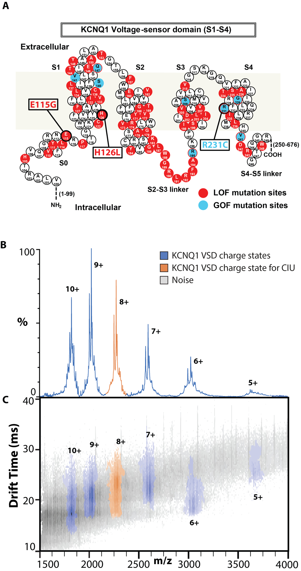
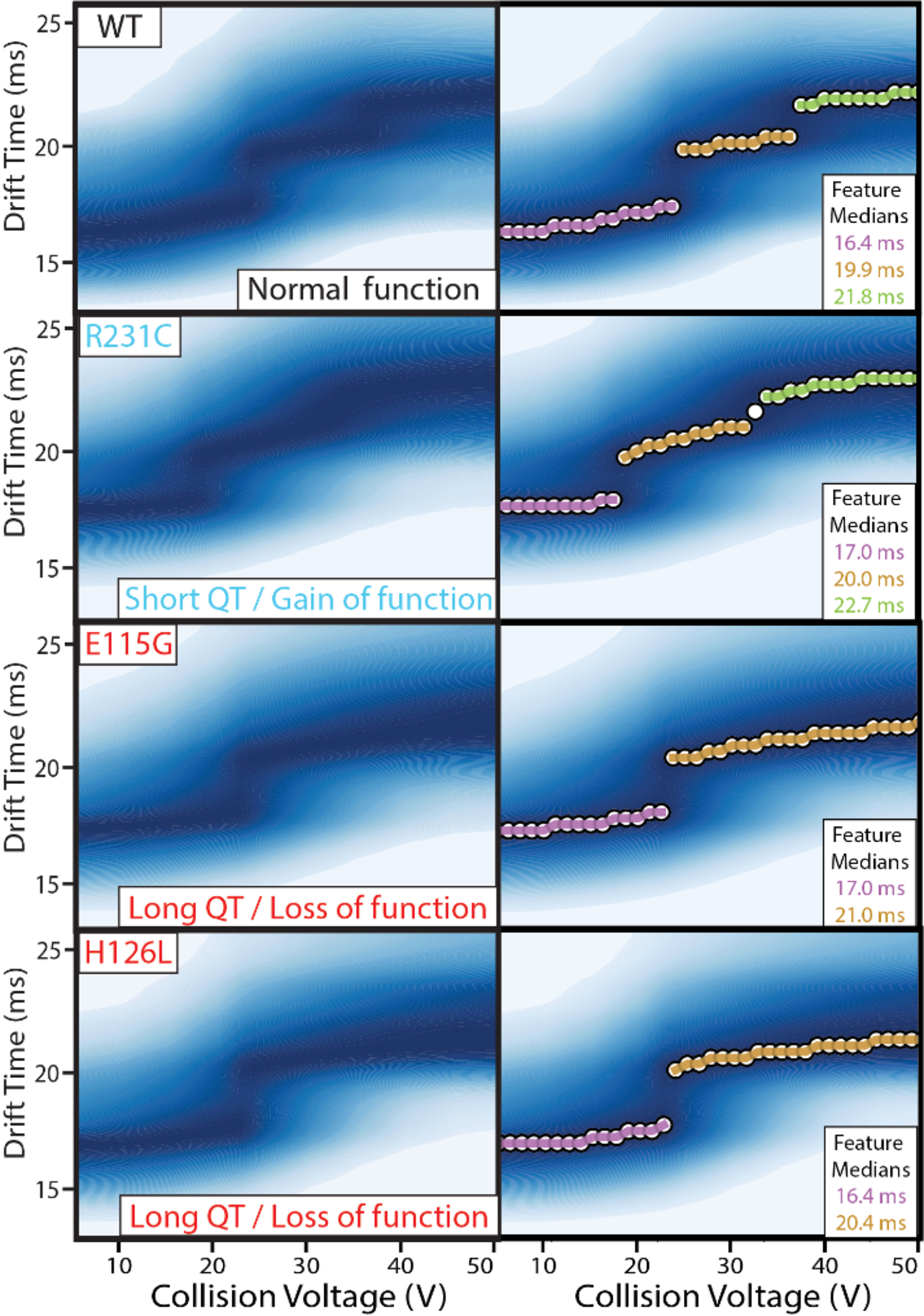
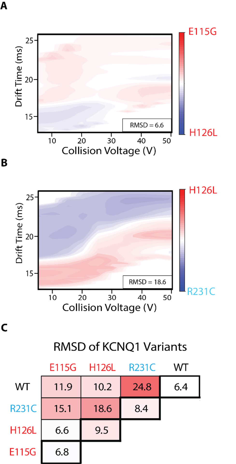
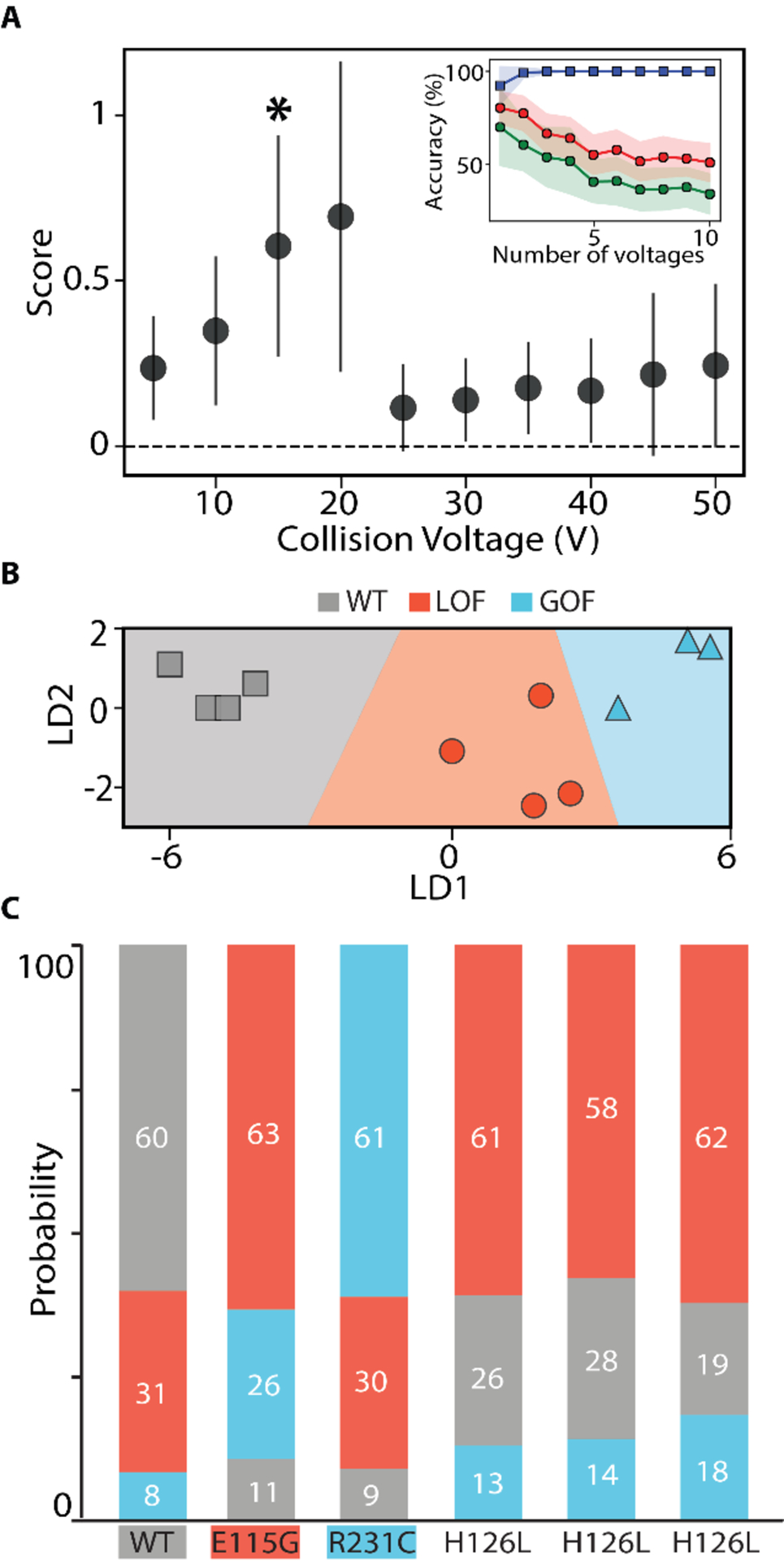
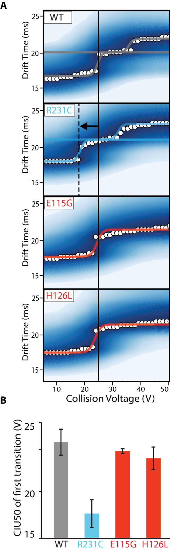
The KCNQ1 voltage-gated potassium channel regulates the repolarization of cardiac cells, and a plurality of point mutations in its voltage-sensing domain (VSD) are associated with toxic gain or loss of pore function, resulting in disease. As is the case with many disease-associated membrane proteins, there are hundreds of human variants of interest identified for KCNQ1; however, a significant portion of these variants have not been characterized in relation to their functional and disease associations. Additionally, as the VSD consists of four transmembrane helices, studies into dynamic structural differences among KCNQ1 VSD variants are hindered by the current limitations and deficits in the high-resolution structure determination of membrane proteins. Here, we use native ion mobility-mass spectrometry and collision-induced unfolding (CIU) to address the need for a high throughput-compatible method for the structural characterization of membrane protein variants of unknown significance using the KCNQ1 VSD as a model system. We perform CIU on wild-type and three mutant KCNQ1 VSD forms associated with the toxic gain or loss of function and show through both automated feature detection and comprehensive difference analysis of the CIU data sets that the variants are clearly grouped by function and disease association. We also construct a classification scheme based on the CIU data sets, which is able to differentiate the variant functional groups and classify a recently characterized variant to its correct grouping. Further, we probe the stability of the KCNQ1 VSD variants when liberated from C12E8 micelles at pH 8.0 and find preliminary evidence that the R231C mutation associated with the gain of the pore function is destabilized relative to the wild-type and loss of function variants.


Conflict of interest statement

The authors declare no competing financial interest.

Figures

Figure 1.
Figure 1.
Native IM-MS of the KCNQ1 VSD. A. Sequence and structure of the KCNQ1 VSD with residues known to mutate and result in disease phenotypes associated with LOF (red) or GOF (blue). Variants E115G, H126L, and R231C are studied in this work. B,C. Representative mass spectra and IM-MS data for WT KCNQ1 at 80 V trap collision voltage. One distribution of charge states, 5–10+, is observed for all variants corresponding to KCNQ1 VSD monomers (Figure S2). The charge state envelope and CCS analysis indicate native like folding, and the 8+ charge state (orange) was chosen for further analysis.
Figure 2.
Figure 2.
CIU fingerprints of KCNQ1 VSD variants. (Top) Fingerprints were collected for the 8+ charge state in triplicate for WT, R231C, E115G, and H126L variants, from 5–50 V, and then denoised and averaged to produce the images shown here. (Bottom) Automated feature detection of the fingerprints finds three discrete features between 5–50 V for WT and R231C, and two discrete features for E115G and H126L. The similarity in starting drift times of the four fingerprints indicate all forms begin at similar orientationally averaged sizes.
Figure 3.
Figure 3.
Comprehensive difference analysis of KCNQ1 VSD variants CIU fingerprints, N = 3 for each variant. A, B. Example difference plots of a low RMSD comparison (E115G and H126L, 6.6%) and high RMSD comparison (H126L and R231C, 18.6%). C. Pairwise comparisons of WT, E115G, H126L, and R231C. RMSD baseline values are shown in the thick bordered boxes. Differences of 2–3x above the baseline are considered significant. All difference plots are shown in Figure S3.
Figure 4.
Figure 4.
CIU based classification of KCNQ1 VSD functional variants A. Each voltage is scored on its ability to differentiate the three classes: WT, LOF, and GOF, using at least three WT, E115G, and R231C replicates. All voltages were used in a ‘leave one out’ cross-validation test, shown in the inset plot, where we tracked the accuracy achieved with the training data (blue),non-training data (green), and the area under the ROC curve (red) as a function of the number of voltages included in the classification scheme. These tests indicated one voltage (*) was best for classification B. Using the voltage indicated in A, the training data set is plotted in linear discriminant and shows clear separation of the data into the three classes. C. The probability of assignment for replicates not part of the training data set is displayed in a bar chart. Each replicate is correctly assigned, including replicates of the H126L variant for which no example was included in the training set.
Figure 5.
Figure 5.
CIU50 stability analysis of KCNQ1 variants. A. Sigmoidal curves are fit between identified features to describe the transition, where WT and R231C have two transitions and E115G and H126L have one transition. B. A bar chart of average CIU50 values extracted from the inflection point determined for each first transition detected, N = 3. Gray is WT, blue is GOF, and red is LOF.

References

    1. Almén MS; Nordström KJV; Fredriksson R; Schiöth HB Mapping the Human Membrane Proteome: A Majority of the Human Membrane Proteins Can Be Classified According to Function and Evolutionary Origin. BMC Biol. 2009, 7, 50. 10.1186/1741-7007-7-50. - DOI - PMC - PubMed
    1. Escribá PV; González-Ros JM; Goñi FM; Kinnunen PKJ; Vigh L; Sánchez-Magraner L; Fernández AM; Busquets X; Horváth I; Barceló-Coblijn G Membranes: A Meeting Point for Lipids, Proteins and Therapies: Translational Medicine. J. Cell. Mol. Med 2008, 12 (3), 829–875. 10.1111/j.1582-4934.2008.00281.x. - DOI - PMC - PubMed
    1. Marinko JT; Huang H; Penn WD; Capra JA; Schlebach JP; Sanders CR Folding and Misfolding of Human Membrane Proteins in Health and Disease: From Single Molecules to Cellular Proteostasis. 2019. 10.1021/acs.chemrev.8b00532. - DOI - PMC - PubMed
    1. Ackerman MJ Genetic Purgatory and the Cardiac Channelopathies: Exposing the Variants of Uncertain/Unknown Significance Issue. Hear. Rhythm 2015, 12 (11), 2325–2331. 10.1016/j.hrthm.2015.07.002. - DOI - PubMed
    1. Oulas A; Minadakis G; Zachariou M; Spyrou GM Selecting Variants of Unknown Significance through Network-Based Gene-Association Significantly Improves Risk Prediction for Disease-Control Cohorts. Sci. Rep 2019, 9 (1), 1–15. 10.1038/s41598-019-39796-w. - DOI - PMC - PubMed

Substances