. 2021 Feb;41(2):253-266.
doi: 10.1177/0271678X20954929. Epub 2020 Sep 22.

Tissue no-reflow despite full recanalization following thrombectomy for anterior circulation stroke with proximal occlusion: A clinical study

Affiliations

Tissue no-reflow despite full recanalization following thrombectomy for anterior circulation stroke with proximal occlusion: A clinical study

Adrien Ter Schiphorst et al. J Cereb Blood Flow Metab. 2021 Feb.

Abstract

Despite early thrombectomy, a sizeable fraction of acute stroke patients with large vessel occlusion have poor outcome. The no-reflow phenomenon, i.e. impaired microvascular reperfusion despite complete recanalization, may contribute to such "futile recanalizations". Although well reported in animal models, no-reflow is still poorly characterized in man. From a large prospective thrombectomy database, we included all patients with intracranial proximal occlusion, complete recanalization (modified thrombolysis in cerebral infarction score 2c-3), and availability of both baseline and 24 h follow-up MRI including arterial spin labeling perfusion mapping. No-reflow was operationally defined as i) hypoperfusion ≥40% relative to contralateral homologous region, assessed with both visual (two independent investigators) and automatic image analysis, and ii) infarction on follow-up MRI. Thirty-three patients were eligible (median age: 70 years, NIHSS: 18, and stroke onset-to-recanalization delay: 208 min). The operational criteria were met in one patient only, consistently with the visual and automatic analyses. This patient recanalized 160 min after stroke onset and had excellent functional outcome. In our cohort of patients with complete and stable recanalization following thrombectomy for intracranial proximal occlusion, severe ipsilateral hypoperfusion on follow-up imaging associated with newly developed infarction was a rare occurrence. Thus, no-reflow may be infrequent in human stroke and may not substantially contribute to futile recanalizations.

Keywords: Perfusion; arterial spin labeling; cerebral blood flow; cerebral infarction diaschisis; large vessel occlusion.


Conflict of interest statement

Declaration of conflicting interests: The author(s) declared no potential conflicts of interest with respect to the research, authorship, and/or publication of this article.

Figures

Figure 1.
Figure 1.
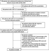
Study flowchart. CCA: common carotid artery; ICA: internal carotid artery; MCA-M1: first segment of middle cerebral artery; MCA-M2: second segment of the middle cerebral artery.
Figure 2.
Figure 2.
Co-registered imaging data for the single patient in whom no-reflow was identified. The patient had sudden-onset severe hemiparesis (three levels) and global aphasia. On admission to our hospital, the neurological deficit had partly resolved and the patient had mild three-level hemiparesis and non-fluent dysphasia (NIHSS = 4). She was treated with thrombectomy 160 min after symptom onset. (a–c) MRI-1 obtained 84 min after symptom onset, showing a small, faint DWI lesion involving the posterior-most lentiform nucleus (a, arrow), an MCA-M1 occlusion on time-of-flight angiogram (b, yellow arrow) distal to the lenticulo-striate arteries (blue arrow), and on PWI (Tmax; the pseudo-color scale ranges from 0 to 10 s) an extensive area of severe hypoperfusion covering a large part of the left cortical MCA territory (yellow arrow), but sparing the deep MCA territory (white arrow) (c). (d) Digital subtraction arteriography obtained immediately following thrombectomy, showing mTICI 3 recanalization. (e–h) Twenty-four-hour MRI-2 showing extension of the DWI lesion to the entire caudate and lentiform nuclei (e), without re-occlusion on time-of-flight MRA (f). These newly infarcted areas are severely hypoperfused on 24 h ASL-CBF maps (g; the pseudo-color scale is in milliliters/100 g/min), fulfilling the operational criteria for no-reflow for both regions (CBF relative to mirror region: –63% and –55%, respectively). No hemorrhage was present across the MCA territory on T2* MRI-2 (h). (a, c, e, g, and h) Images co-registered using GE Volume Viewer (see Methods). Stroke etiology was atrial fibrillation. This patient enjoyed early neurological recovery (24-h NIHSS = 1) and excellent outcome (three-month mRS = 0).
Figure 3.
Figure 3.
Illustrative image dataset of the patient with mild hypoperfusion associated with infarction. This 66-year-old patient presented with acute-onset moderate deficit (NIHSS score = 11) and was treated with IV thrombolysis followed by thrombectomy for right terminal ICA occlusion, with mTICI 2c recanalization obtained 215 min after symptom onset. Perfusion MRI was carried out prior to thrombolysis in this patient but unfortunately the data were not interpretable due to marked head motion and technical errors. (a–c) Illustrative DWI b = 1000 s/mm2 sequences obtained (i) on admission (68 min after symptom onset) (a), and at 24 h follow-up (b and c), showing a stable DWI lesion in the striato-capsular area (i.e. deep MCA territory); (d–f) 24 h follow-up ASL-CBF maps, showing mild hypoperfusion within the DWI lesion (15% and 17% reduction in the caudate and lenticular nuclei, respectively, relative to contralateral ASPECTS regions; white arrows), but also cortically at distance from the DWI lesion (20–35% reduction relative to contralateral ASPECTS regions; red arrows), spreading beyond the ICA territory (i.e. posterior circulation, yellow arrows; note that in this patient both posterior cerebral arteries originated from the basilar artery on MRA, data not shown). The left cerebellar hemisphere, i.e. contralateral to the stroke, also shows mild hypoperfusion compared to the right cerebellum (purple arrow), reflecting crossed cerebellar diaschisis. The patient made a good recovery (24 h NIHSS score = 1, three-month mRS = 1).

Similar articles

Cited by

References

    1. Goyal M, Menon BK, van Zwam WH, et al.. Endovascular thrombectomy after large-vessel ischaemic stroke: a meta-analysis of individual patient data from five randomised trials. Lancet 2016; 387: 1723–1731. - PubMed
    1. Molina CA.Futile recanalization in mechanical embolectomy trials: a call to improve selection of patients for revascularization. Stroke 2010; 41: 842–843. - PubMed
    1. Savitz SI, Baron JC, Yenari MA, et al.. Reconsidering neuroprotection in the reperfusion era. Stroke 2017; 48: 3413–3419. - PubMed
    1. Baron J-C.Protecting the ischaemic penumbra as an adjunct to thrombectomy for acute stroke. Nat Rev Neurol 2018; 14: 325–337. - PubMed
    1. De Silva DA, Fink JN, Christensen S, et al.. Assessing reperfusion and recanalization as markers of clinical outcomes after intravenous thrombolysis in the echoplanar imaging thrombolytic evaluation trial (EPITHET). Stroke 2009; 40: 2872–2874. - PubMed

Publication types