

Reorganization of the nuclear architecture in the Drosophila melanogaster Lamin B mutant lacking the CaaX box
- PMID: 32960740
- PMCID: PMC7529411
- DOI: 10.1080/19491034.2020.1819704
Reorganization of the nuclear architecture in the Drosophila melanogaster Lamin B mutant lacking the CaaX box
Abstract
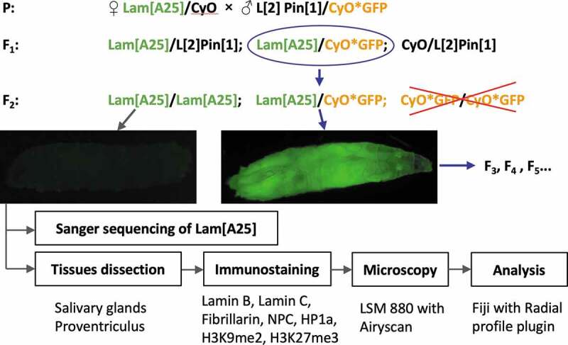
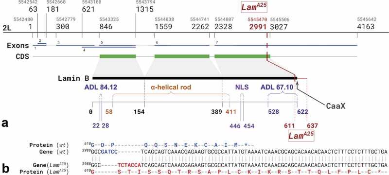
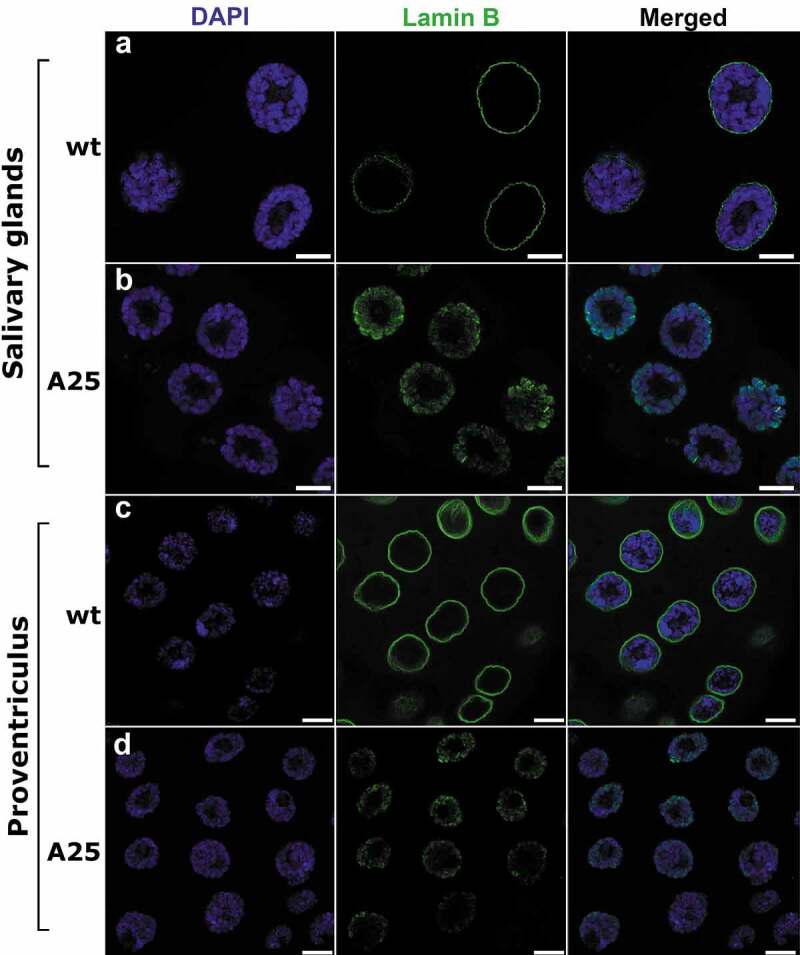
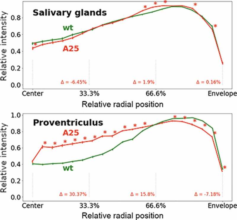
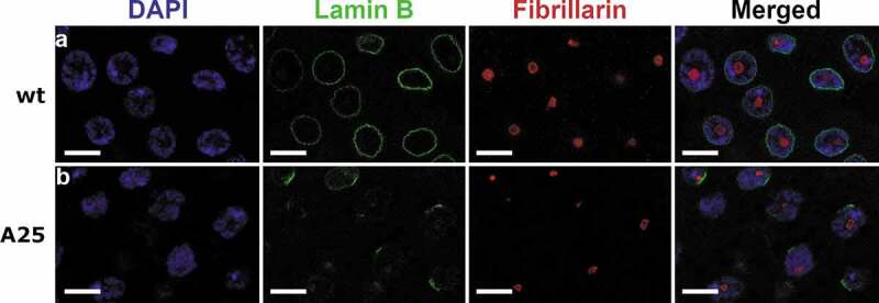
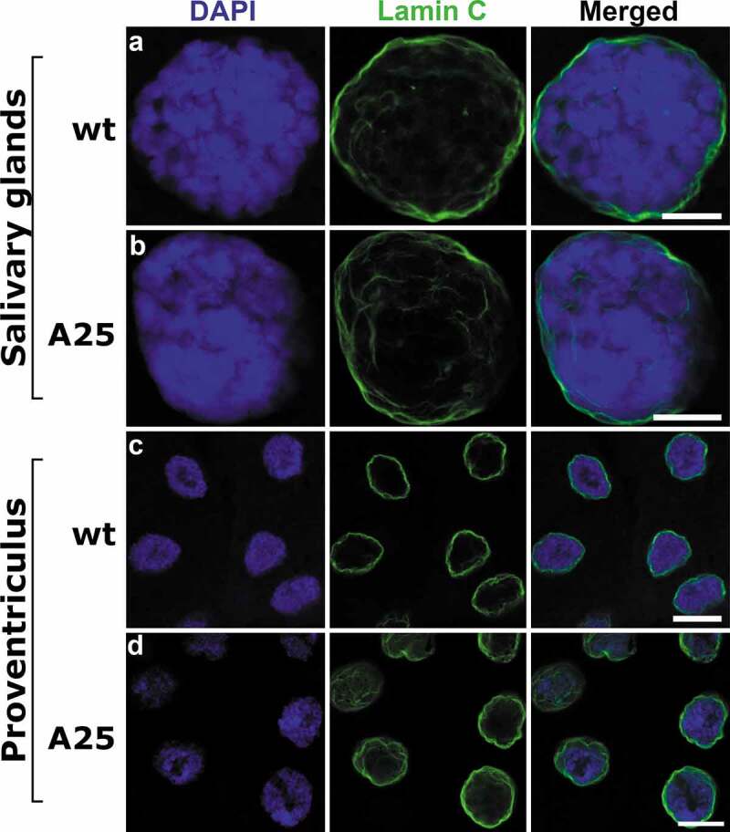
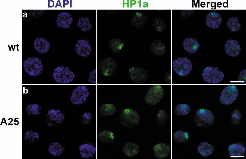
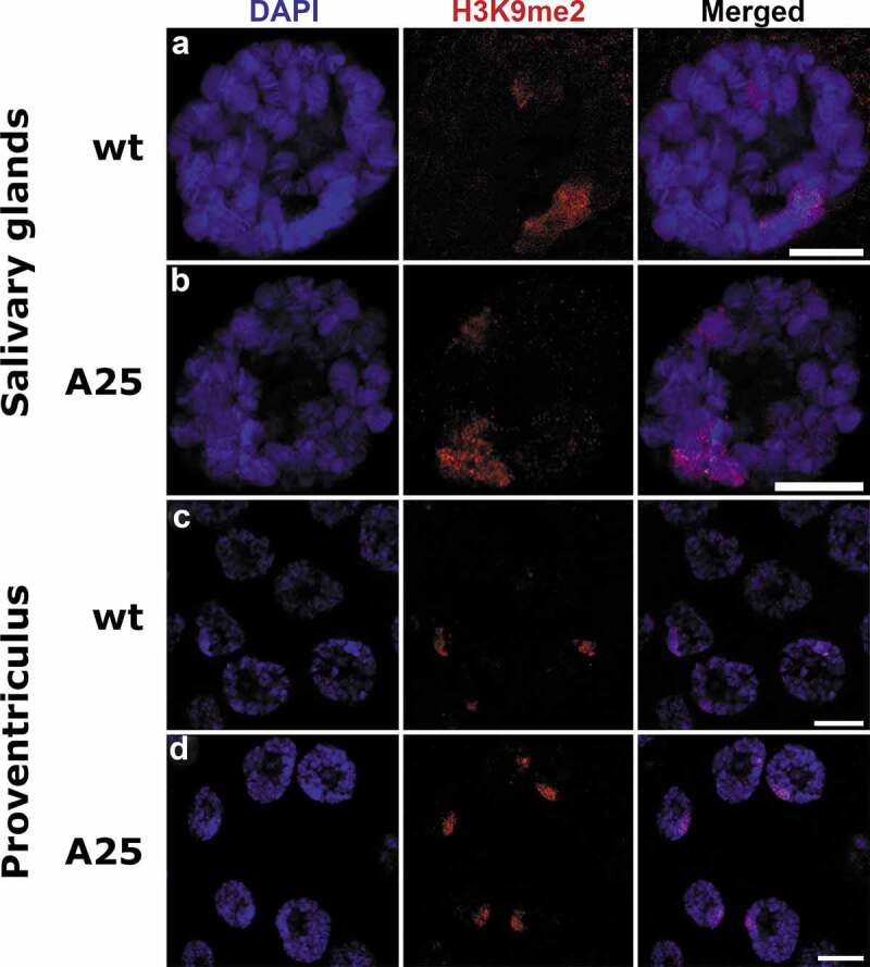
Lamins interact with the nuclear membrane and chromatin but the precise players and mechanisms of these interactions are unknown. Here, we tested whether the removal of the CaaX motif from Lamin B disrupts its attachment to the nuclear membrane and affects chromatin distribution. We usedDrosophila melanogaster LamA25 homozygous mutants that lack the CaaX box. We found that the mutant Lamin B was not confined to the nuclear periphery but was distributed throughout the nuclear interior, colocalizing with chromosomes in salivary gland and proventriculus. The peripheral position of Lamin C, nuclear pore complex (NPC), heterochromatin protein 1a (HP1a), H3K9me2- and H3K27me3-associated chromatin remained intact. The fluorescence intensity of the DAPI-stained peripheral chromatin significantly decreased and that of the central chromatin significantly increased in the proventriculus nuclei of the mutantflies compared to wild-type. However, the mutation had little effect on chromatin radial distribution inside highly polytenized salivary gland nuclei.
Keywords: Dm0; Drosophila; Lama25 mutant; B-type lamin; Lamin B; Nuclear lamina; chromatin; confocal microscopy; nuclear envelope; proventriculus nuclei; salivary gland nuclei.
Conflict of interest statement
No potential conflict of interest is reported by the authors.
Figures

Similar articles
-
Specific and conserved sequences in D. melanogaster and C. elegans lamins and histone H2A mediate the attachment of lamins to chromosomes.J Cell Sci. 2007 Jan 1;120(Pt 1):77-85. doi: 10.1242/jcs.03325. Epub 2006 Dec 5. J Cell Sci. 2007. PMID: 17148572
-
Non-farnesylated B-type lamin can tether chromatin inside the nucleus and its chromatin interaction requires the Ig-fold region.Chromosoma. 2017 Feb;126(1):125-144. doi: 10.1007/s00412-016-0581-x. Epub 2016 Feb 19. Chromosoma. 2017. PMID: 26892013
-
The large fraction of heterochromatin in Drosophila neurons is bound by both B-type lamin and HP1a.Epigenetics Chromatin. 2018 Nov 1;11(1):65. doi: 10.1186/s13072-018-0235-8. Epigenetics Chromatin. 2018. PMID: 30384843 Free PMC article.
-
Lamins and lamin-binding proteins in functional chromatin organization.Crit Rev Eukaryot Gene Expr. 1999;9(3-4):257-65. doi: 10.1615/critreveukargeneexpr.v9.i3-4.100. Crit Rev Eukaryot Gene Expr. 1999. PMID: 10651242 Review.
-
Nuclear Lamins: Thin Filaments with Major Functions.Trends Cell Biol. 2018 Jan;28(1):34-45. doi: 10.1016/j.tcb.2017.08.004. Epub 2017 Sep 8. Trends Cell Biol. 2018. PMID: 28893461 Review.
Cited by
-
Effects of Lamina-Chromatin Attachment on Super Long-Range Chromatin Interactions.bioRxiv [Preprint]. 2025 Feb 17:2025.02.13.638183. doi: 10.1101/2025.02.13.638183. bioRxiv. 2025. PMID: 40027763 Free PMC article. Preprint.
-
The probability of chromatin to be at the nuclear lamina has no systematic effect on its transcription level in fruit flies.Epigenetics Chromatin. 2024 May 6;17(1):13. doi: 10.1186/s13072-024-00528-8. Epigenetics Chromatin. 2024. PMID: 38705995 Free PMC article.
-
Quantifying conformational heterogeneity of 3D genome organization in fruit fly.PLoS One. 2025 Jul 3;20(7):e0326927. doi: 10.1371/journal.pone.0326927. eCollection 2025. PLoS One. 2025. PMID: 40608785 Free PMC article.
-
Four-Dimensional Mesoscale Liquid Model of Nucleus Resolves Chromatin's Radial Organization.PRX Life. 2024 Jan-Mar;2(1):013006. doi: 10.1103/PRXLife.2.013006. Epub 2024 Jan 30. PRX Life. 2024. PMID: 38601142 Free PMC article.
-
Quantifying Conformational Heterogeneity of 3D Genome Organization in Fruit Fly.bioRxiv [Preprint]. 2025 May 27:2025.05.24.655945. doi: 10.1101/2025.05.24.655945. bioRxiv. 2025. Update in: PLoS One. 2025 Jul 3;20(7):e0326927. doi: 10.1371/journal.pone.0326927. PMID: 40502076 Free PMC article. Updated. Preprint.
References
-
- Stuurman N, Heins S, Aebi U. Nuclear lamins: their structure, assembly, and interactions. J Struct Biol. 1998;122:42–66. - PubMed
-
- Bao X, Girton J, Johansen J, et al. The lamin Dm0 allele Ari3 acts as an enhancer of position effect variegation of the wm4 allele in Drosophila. Genetica. 2007;129:339–342. - PubMed
-
- de Leeuw R, Gruenbaum Y, Nuclear Lamins MO. Thin filaments with major functions. Trends Cell Biol. 2018;28:34–45. - PubMed
Publication types
MeSH terms
Substances
LinkOut - more resources
Full Text Sources
Other Literature Sources
Molecular Biology Databases