Case Reports
. 2021 Jan 4;131(1):e139980.
doi: 10.1172/JCI139980.

Herpes simplex encephalitis in a patient with a distinctive form of inherited IFNAR1 deficiency

Affiliations
Case Reports

Herpes simplex encephalitis in a patient with a distinctive form of inherited IFNAR1 deficiency

Paul Bastard et al. J Clin Invest. .

Abstract

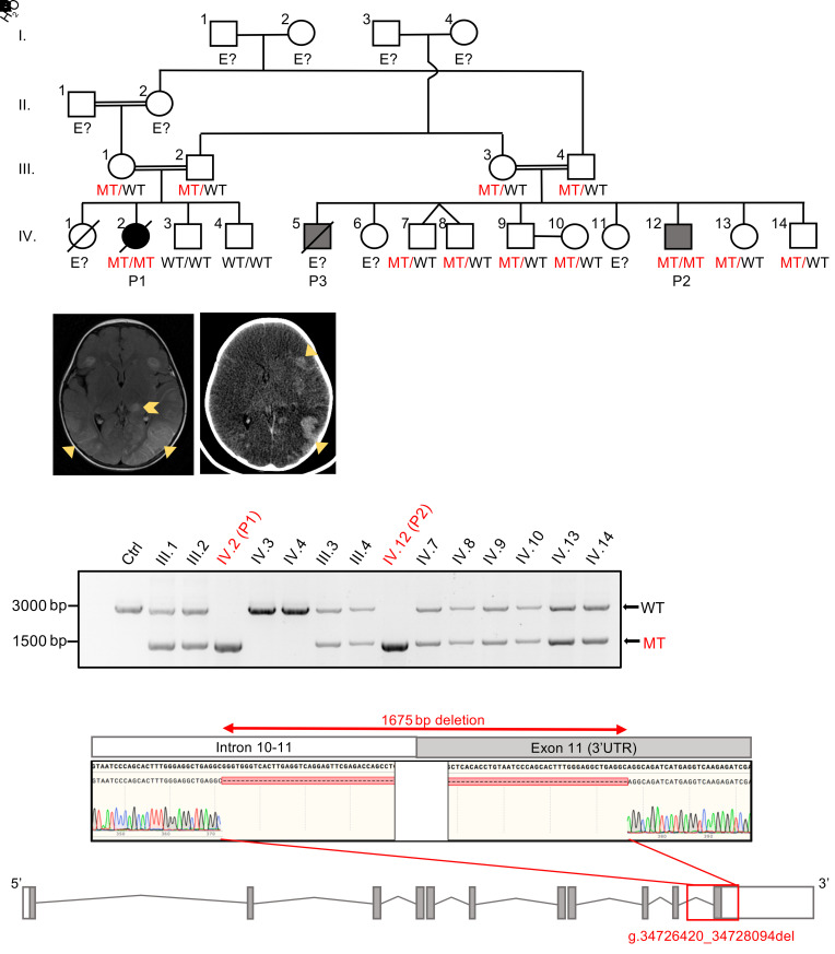
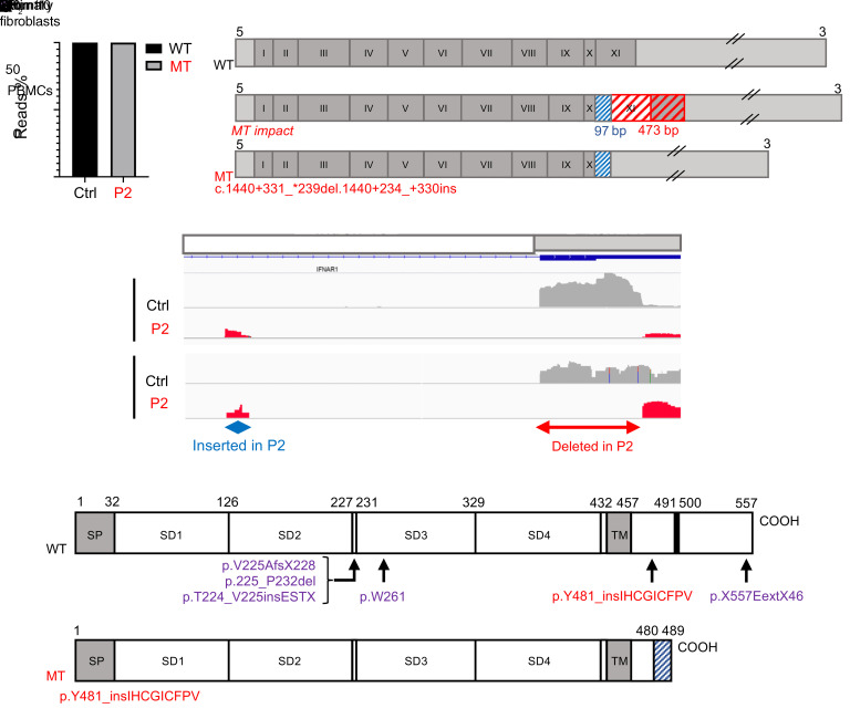
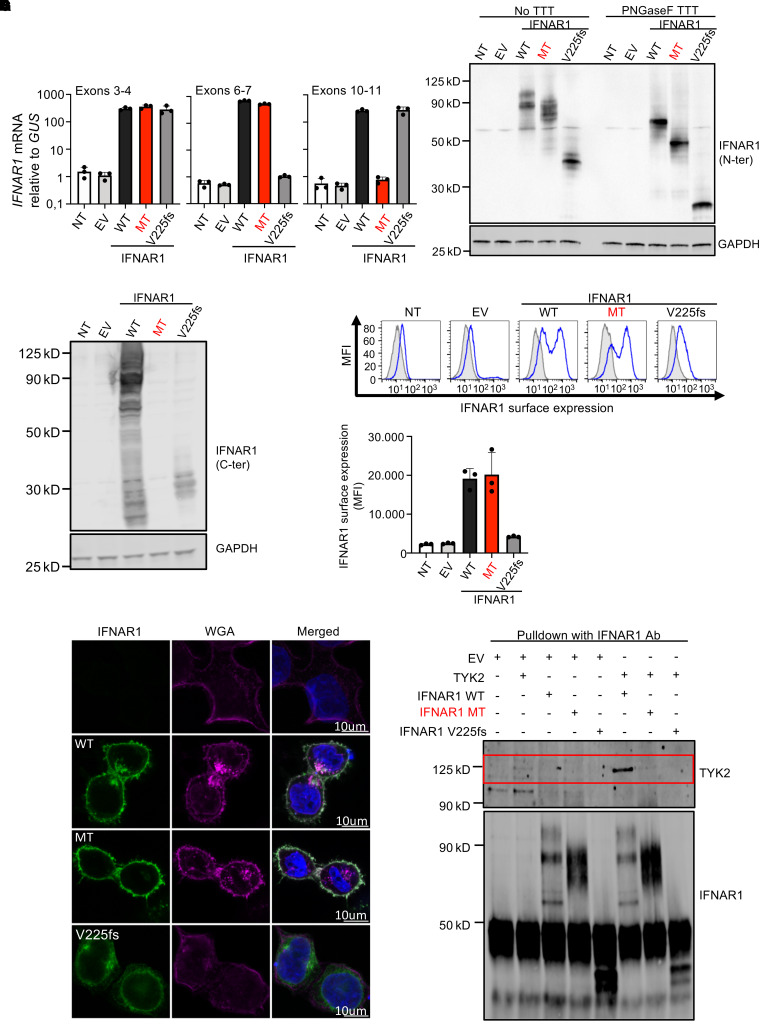
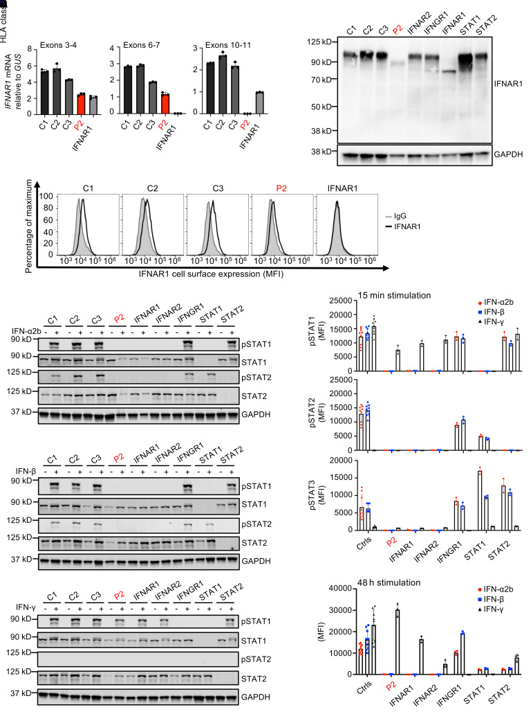
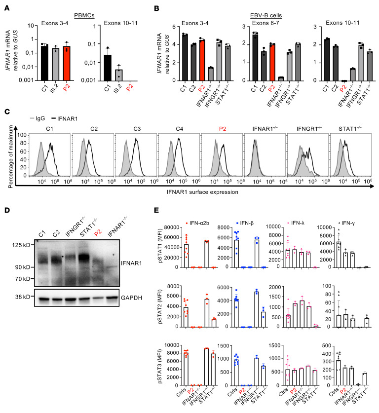
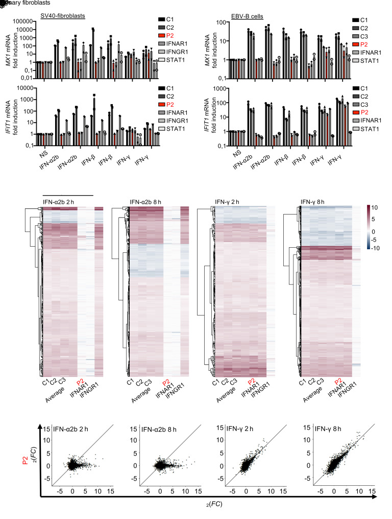
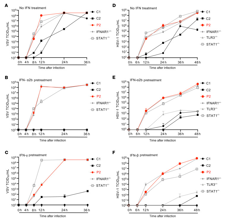
Inborn errors of TLR3-dependent IFN-α/β- and IFN-λ-mediated immunity in the CNS can underlie herpes simplex virus 1 (HSV-1) encephalitis (HSE). The respective contributions of IFN-α/β and IFN-λ are unknown. We report a child homozygous for a genomic deletion of the entire coding sequence and part of the 3'-UTR of the last exon of IFNAR1, who died of HSE at the age of 2 years. An older cousin died following vaccination against measles, mumps, and rubella at 12 months of age, and another 17-year-old cousin homozygous for the same variant has had other, less severe, viral illnesses. The encoded IFNAR1 protein is expressed on the cell surface but is truncated and cannot interact with the tyrosine kinase TYK2. The patient's fibroblasts and EBV-B cells did not respond to IFN-α2b or IFN-β, in terms of STAT1, STAT2, and STAT3 phosphorylation or the genome-wide induction of IFN-stimulated genes. The patient's fibroblasts were susceptible to viruses, including HSV-1, even in the presence of exogenous IFN-α2b or IFN-β. HSE is therefore a consequence of inherited complete IFNAR1 deficiency. This viral disease occurred in natural conditions, unlike those previously reported in other patients with IFNAR1 or IFNAR2 deficiency. This experiment of nature indicates that IFN-α/β are essential for anti-HSV-1 immunity in the CNS.

Keywords: Cytokines; Genetic diseases; Immunology; Infectious disease; Innate immunity.


Conflict of interest statement

Conflict of interest: The authors have declared that no conflict of interest exists.

Figures

Figure 1
Figure 1. Homozygous deletion in IFNAR1 in a patient who died from HSE and her cousin.
(A) Family pedigree showing the segregation of the IFNAR1 mutant (MT) allele. Double lines connect the 2 parents with consanguinity. The filled black symbol indicates the proband (patient 1, P1) with HSE, the filled gray symbols indicate individuals with viral diseases other than HSE, and the open symbols indicate healthy family members. E?, unknown IFNAR1 genotype. (B) Brain imaging showing HSE lesions in P1. Left: Post-contrast T2-FLAIR image showing diffuse cortical and subcortical edema on temporo-occipital regions accompanied by leptomeningeal enhancement (yellow triangles), compatible with meningoencephalitis. There are also parenchymal lesions, involving the left thalamus and base of the frontal lobes, indicated by yellow arrowhead. Right: Noncontrast head CT, performed 7 days later, showing diffuse brain edema with multiple parenchymal hemorrhages in the edematous areas previously identified (yellow triangles). (C) IFNAR1 from leukocyte gDNA from the patients and other relatives, amplified by PCR with a forward primer binding to exon 10 and a reverse primer binding to the 3′-UTR part of exon 11. The result shown is representative of 2 independent experiments. (D) Top: Sanger sequencing results for IFNAR1 from patient leukocyte gDNA. Bottom: Schematic diagram of the IFNAR1 gene in the gDNA, with 11 coding exons, and a red box representing the location of the deletion found in the patients. The result shown is representative of 3 independent experiments on 2 independently drawn samples.
Figure 2
Figure 2. The IFNAR1 deletion leads to aberrant cDNA splicing.
(A) cDNA TOPO cloning and sequencing results demonstrating complete aberrant splicing of IFNAR1 in PBMCs from P2. At least 100 transcripts were sequenced for the patient and the control. The result shown is the sum of 2 independent experiments. (B) Schematic diagram of the full-length cDNA of WT and MT IFNAR1. Sequencing results demonstrated aberrant splicing and an absence of the coding sequence of IFNAR1 exon 11 in PBMCs from P2. The blue box indicates the 97-bp intronic insertion. The red box indicates the 473-bp deletion. The exons are numbered in roman numerals (I–XI). The 5′- or 3′-UTR is shown in light gray, and the coding sequences of the exons are shown in dark gray. (C) RNA-Seq results for primary fibroblasts (top) or PBMCs (bottom) showing the coverage of IFNAR1 from intron 10 to exon 11, in healthy controls and P2, demonstrating an insertion and the deletion of the coding sequence of exon 11 and part of the 3′-UTR. For primary fibroblasts, the results shown are representative of 5 technical replicates (corresponding to different RNA-Seq conditions). (D) Schematic diagram of the WT and MT IFNAR1 proteins, with the 4 fibronectin type III subdomains (SD1–SD4) and the TYK2 interaction domain (in black). The signal peptide is denoted “SP” and the transmembrane domain “TM.” The mutation reported in this study is indicated in red, and previously reported mutations are indicated in violet.
Figure 3
Figure 3. The MT IFNAR1 protein is expressed on the cell surface, truncated, and does not bind TYK2.
(A) Real-time qPCR for IFNAR1 in HEK293T cells transiently transfected with IFNAR1 cDNA constructs; GUS was used as an expression control. Mean value and SD from 3 independent experiments with technical duplicates in each experiment. (B) Western blot (WB) of IFNAR1 in HEK293T cells transiently transfected with IFNAR1 cDNA constructs, and the same samples treated with PNGase F to inhibit glycosylation. An antibody recognizing the N-terminal (N-ter) part of the IFNAR1 protein was used. GAPDH was used as a loading control. A representative blot from 3 independent experiments is shown. NT, nontransfected; EV, empty vector; V225fs, variant of the previously reported IFNAR1–/– patient. TTT, treatment. (C) WB of IFNAR1 in HEK293T cells transiently transfected with IFNAR1 constructs. An antibody recognizing the C-terminal (C-ter) part of the protein was used. GAPDH was used as a loading control. A representative blot from 2 independent experiments is shown. (D) Extracellular FACS staining and mean fluorescence intensity (MFI) of IFNAR1 in HEK cells transiently transfected with IFNAR1 cDNA constructs, with an antibody recognizing the N-terminus of the protein. Cells were not permeabilized. Results representative of 3 independent experiments are shown. (E) MFI of IFNAR1 surface expression, represented graphically. Mean values and SD from 3 independent experiments are shown. (F) Immunofluorescence staining, as assessed by confocal microscopy in HeLa cells transiently transfected with IFNAR1 cDNA constructs. An antibody against the N-terminus of IFNAR1 was used (green), and membranes were stained for wheat germ agglutinin (WGA) (purple). The nuclei were stained with DAPI (blue). The images shown are representative of 3 independent experiments. (G) WB for TYK2 and IFNAR1 after coimmunoprecipitation from protein extracts of HEK293T cells cotransfected with WT or MT IFNAR1 cDNA constructs and WT TYK2. The images presented are representative of 3 independent experiments.
Figure 4
Figure 4. Patient SV40-fibroblasts express a truncated IFNAR1 and do not respond to IFN-α/β.
(A) IFNAR1 mRNA levels in SV40-fibroblasts from 3 healthy controls (C1, C2, C3), P2, and the previously reported p.V225fs IFNAR1–/– patient; GUS was used as an expression control. Mean values and SD from 3 independent experiments, each with technical duplicates, are shown. (B) WB for IFNAR1 in SV40-fibroblasts from 3 healthy controls (C1, C2, C3), P2, and other patients with autosomal recessive (AR) complete deficiencies of the IFN signaling pathways (IFNAR1–/–, IFNAR2–/–, IFNGR1–/–, STAT1–/–, STAT2–/–). A truncated form of IFNAR1 was observed in the cells from the previously reported IFNAR1–/– patient, as indicated by the blue asterisk. An antibody recognizing the N-terminal part of the IFNAR1 protein was used. GAPDH was used as a loading control. (C) Extracellular FACS staining of IFNAR1 in SV40-fibroblasts from 3 healthy controls (C1, C2, C3), P2, and the previously reported IFNAR1–/– patient. Cells were not permeabilized. An antibody recognizing the N-terminal part of the protein was used. (D) WB of p-STAT1, p-STAT2, and unphosphorylated STAT1 and STAT2 in SV40-fibroblasts stimulated with 1000 U/mL IFN-α2b, IFN-β, or IFN-γ for 15 minutes. The cells used were from 3 healthy controls (C1, C2, C3), P2, and IFNAR1–/–, IFNAR2–/–, IFNGR1–/–, STAT1–/–, and STAT2–/– patients. GAPDH was used as a loading control. The results shown in BD are representative of 3 independent experiments. (E) MFI following the intracellular FACS staining of p-STAT1, p-STAT2, and p-STAT3 in SV40-fibroblasts stimulated with 1000 U/mL IFN-α2b, IFN-β, or IFN-γ for 15 minutes. The values presented are after subtraction of the nonstimulated condition’s value. (F) MFI after extracellular FACS staining of HLA class I in SV40-fibroblasts stimulated with 1000 U/mL IFN-α2b, IFN-β, or IFN-γ for 48 hours. MFI (and SD) values from 3 independent experiments are shown in E and F.
Figure 5
Figure 5. Patient EBV-B cells express a truncated IFNAR1 and do not respond to IFN-α/β.
(A) IFNAR1 mRNA levels, in PBMCs from 1 healthy control, P1’s father (III.2), and P2. The GUS housekeeping gene was used as an expression control. Mean (and SD) values from 3 independent experiments, each with technical duplicates, are shown. (B) IFNAR1 mRNA levels in EBV-B cells from 2 healthy controls, P2, and previously reported IFNAR1–/–, IFNGR1–/–, and STAT1–/– patients; the housekeeping gene GUS was used as an expression control. Mean (and SD) values from 3 independent experiments, each with technical duplicates, are shown. (C) Extracellular FACS staining of IFNAR1 in EBV-B cells from 4 healthy controls, P2, and IFNAR1–/–, IFNGR1–/–, and STAT1–/– patients. Cells were not permeabilized. An antibody recognizing the N-terminal part of the protein was used. The results shown are representative of 3 independent experiments. (D) WB of IFNAR1 in EBV-B cells from 2 healthy controls (C1, C2), P2, and IFNAR1–/–, IFNGR1–/–, and STAT1–/– patients. An antibody recognizing the N-terminal side of the protein was used. GAPDH was used as a loading control. A representative blot from 3 independent experiments is shown. (E) MFI after intracellular FACS staining of p-STAT1, p-STAT2, and p-STAT3, in EBV-B cells stimulated with 1000 U/mL IFN-α2b, IFN-β, IFN-λ, or IFN-γ for 15 minutes. The cells used were from 3 healthy controls (C1, C2, C3), P2, and IFNAR1–/–, IFNGR1–/–, and STAT1–/– patients. MFI (and SD) values from 3 independent experiments are shown.
Figure 6
Figure 6. Abolition of the induction of ISGs in response to IFN-α/β in patient fibroblasts and EBV-B cells.
(A) Fold change in IFIT1 and MX1 mRNA levels after the stimulation, with IFN-α2b, IFN-β, or IFN-γ, of SV40-fibroblasts from 2 healthy controls (C1, C2), P2, and IFNAR1–/–, IFNGR1–/–, and STAT1–/– patients for 2 hours or 8 hours. The GUS housekeeping gene was used as an expression control. Mean and SD values from 3 independent experiments are shown. (B) Fold change in IFIT1 and MX1 mRNA levels after the stimulation, with IFN-α2b, IFN-β, or IFN-γ, of EBV-B cells from 3 healthy controls (C1, C2, C3), P2, and the IFNAR1–/– and STAT1–/– patients for 2 hours or 8 hours. The housekeeping gene GUS was used as an expression control. Mean and SD values from 3 independent experiments are shown. (C) Heatmaps of genes differentially expressed after 2 or 8 hours of stimulation with IFN‑α2b or IFN‑γ, in primary fibroblasts from 3 healthy controls (C1, C2, C3), P2, the IFNAR1–/– p.V225fs patient, and an IFNGR1–/– patient. For each set of conditions, we show the genes significantly differentially expressed with respect to the control group, i.e., with a |log2(fold change [FC])| >1 and P < 0.05 after Benjamini-Hochberg correction, and genes with a |Δlog2(FC)| >1 between the control group and the IFNAR1–/– patient or the IFNGR1–/– patient after stimulation with IFN-α2b and IFN-γ, respectively. The gradient from blue to red represents increasing log2(FC). Genes are clustered by Euclidean distance. (D) Scatterplots of log2(FC) in RNA-Seq–quantified gene expression following stimulation with IFN‑α2b or IFN‑γ for 2 or 8 hours, in primary fibroblasts from P2 versus 3 healthy controls (C1, C2, and C3). Each dot represents a single gene.
Figure 7
Figure 7. Enhanced viral replication in patient SV40-fibroblasts in the presence and absence of treatment with IFN-α2b or IFN-β.
(AC) VSV replication, quantified by the TCID50 method, in SV40-fibroblasts from 2 healthy controls (C1, C2), P2, and IFNAR1–/– and STAT1–/– patients, 4, 8, 12, 24, and 36 hours after infection with VSV at an MOI of 3, without (A) or with prior treatment with 1000 IU IFN-α2b (B) or IFN-γ (C) for 16 hours. Mean values ± SD from 3 independent experiments, each including biological triplicates, are shown. (DF) HSV-1 replication, quantified by the TCID50 method, in SV40-fibroblasts from 2 healthy controls, P2, and IFNAR1–/–, TLR3–/–, and STAT1–/– patients, 6, 12, 24, 36, and 48 hours after infection with HSV-1 at an MOI of 1, without (D) or with prior treatment with 1000 IU IFN-α2b (E) or IFN-β (F) for 16 hours. Mean values ± SD from 3 independent experiments are shown (AF). Biological triplicates were included in each experiment for VSV infection, with duplicates for HSV-1 infection.

Similar articles

  • Heterozygous TBK1 mutations impair TLR3 immunity and underlie herpes simplex encephalitis of childhood.
    Herman M, Ciancanelli M, Ou YH, Lorenzo L, Klaudel-Dreszler M, Pauwels E, Sancho-Shimizu V, Pérez de Diego R, Abhyankar A, Israelsson E, Guo Y, Cardon A, Rozenberg F, Lebon P, Tardieu M, Heropolitanska-Pliszka E, Chaussabel D, White MA, Abel L, Zhang SY, Casanova JL. Herman M, et al. J Exp Med. 2012 Aug 27;209(9):1567-82. doi: 10.1084/jem.20111316. Epub 2012 Jul 30. J Exp Med. 2012. PMID: 22851595 Free PMC article.
  • Impaired intrinsic immunity to HSV-1 in human iPSC-derived TLR3-deficient CNS cells.
    Lafaille FG, Pessach IM, Zhang SY, Ciancanelli MJ, Herman M, Abhyankar A, Ying SW, Keros S, Goldstein PA, Mostoslavsky G, Ordovas-Montanes J, Jouanguy E, Plancoulaine S, Tu E, Elkabetz Y, Al-Muhsen S, Tardieu M, Schlaeger TM, Daley GQ, Abel L, Casanova JL, Studer L, Notarangelo LD. Lafaille FG, et al. Nature. 2012 Nov 29;491(7426):769-73. doi: 10.1038/nature11583. Epub 2012 Oct 28. Nature. 2012. PMID: 23103873 Free PMC article.
  • Genetic defects of brain immunity in childhood herpes simplex encephalitis.
    Zhang SY, Casanova JL. Zhang SY, et al. Nature. 2024 Nov;635(8039):563-573. doi: 10.1038/s41586-024-08119-z. Epub 2024 Nov 20. Nature. 2024. PMID: 39567785 Free PMC article. Review.
  • Human SNORA31 variations impair cortical neuron-intrinsic immunity to HSV-1 and underlie herpes simplex encephalitis.
    Lafaille FG, Harschnitz O, Lee YS, Zhang P, Hasek ML, Kerner G, Itan Y, Ewaleifoh O, Rapaport F, Carlile TM, Carter-Timofte ME, Paquet D, Dobbs K, Zimmer B, Gao D, Rojas-Duran MF, Kwart D, Rattina V, Ciancanelli MJ, McAlpine JL, Lorenzo L, Boucherit S, Rozenberg F, Halwani R, Henry B, Amenzoui N, Alsum Z, Marques L, Church JA, Al-Muhsen S, Tardieu M, Bousfiha AA, Paludan SR, Mogensen TH, Quintana-Murci L, Tessier-Lavigne M, Smith GA, Notarangelo LD, Studer L, Gilbert W, Abel L, Casanova JL, Zhang SY. Lafaille FG, et al. Nat Med. 2019 Dec;25(12):1873-1884. doi: 10.1038/s41591-019-0672-3. Epub 2019 Dec 5. Nat Med. 2019. PMID: 31806906 Free PMC article.
  • Mendelian predisposition to herpes simplex encephalitis.
    Zhang SY, Abel L, Casanova JL. Zhang SY, et al. Handb Clin Neurol. 2013;112:1091-7. doi: 10.1016/B978-0-444-52910-7.00027-1. Handb Clin Neurol. 2013. PMID: 23622315 Review.

Cited by

  • Immunity and Genetics at the Revolving Doors of Diagnostics in Primary Immunodeficiencies.
    Rispoli F, Valencic E, Girardelli M, Pin A, Tesser A, Piscianz E, Boz V, Faletra F, Severini GM, Taddio A, Tommasini A. Rispoli F, et al. Diagnostics (Basel). 2021 Mar 16;11(3):532. doi: 10.3390/diagnostics11030532. Diagnostics (Basel). 2021. PMID: 33809703 Free PMC article.
  • SARS-CoV-2 brainstem encephalitis in human inherited DBR1 deficiency.
    Chan YH, Lundberg V, Le Pen J, Yuan J, Lee D, Pinci F, Volpi S, Nakajima K, Bondet V, Åkesson S, Khobrekar NV, Bodansky A, Du L, Melander T, Mariaggi AA, Seeleuthner Y, Saleh TS, Chakravarty D, Marits P, Dobbs K, Vonlanthen S, Hennings V, Thörn K, Rinchai D, Bizien L, Chaldebas M, Sobh A, Özçelik T, Keles S, AlKhater SA, Prando C, Meyts I; COVID Human Genetic Effort; Wilson MR, Rosain J, Jouanguy E, Aubart M, Abel L, Mogensen TH, Pan-Hammarström Q, Gao D, Duffy D, Cobat A, Berg S, Notarangelo LD, Harschnitz O, Rice CM, Studer L, Casanova JL, Ekwall O, Zhang SY. Chan YH, et al. J Exp Med. 2024 Sep 2;221(9):e20231725. doi: 10.1084/jem.20231725. Epub 2024 Jul 18. J Exp Med. 2024. PMID: 39023559 Free PMC article.
  • Human autoantibodies neutralizing type I IFNs: From 1981 to 2023.
    Bastard P, Gervais A, Le Voyer T, Philippot Q, Cobat A, Rosain J, Jouanguy E, Abel L, Zhang SY, Zhang Q, Puel A, Casanova JL. Bastard P, et al. Immunol Rev. 2024 Mar;322(1):98-112. doi: 10.1111/imr.13304. Epub 2024 Jan 9. Immunol Rev. 2024. PMID: 38193358 Free PMC article. Review.
  • From a focal skin issue to a systemic disease: the multifaceted nature of cold sores, novel findings.
    Akhoundi N, Noorbakhsh M, Siami A, Naseri Z, Hazara S, Hazara K. Akhoundi N, et al. Virusdisease. 2024 Sep;35(3):428-433. doi: 10.1007/s13337-024-00877-8. Epub 2024 Jul 9. Virusdisease. 2024. PMID: 39464735
  • Impaired IL-23-dependent induction of IFN-γ underlies mycobacterial disease in patients with inherited TYK2 deficiency.
    Ogishi M, Arias AA, Yang R, Han JE, Zhang P, Rinchai D, Halpern J, Mulwa J, Keating N, Chrabieh M, Lainé C, Seeleuthner Y, Ramírez-Alejo N, Nekooie-Marnany N, Guennoun A, Muller-Fleckenstein I, Fleckenstein B, Kilic SS, Minegishi Y, Ehl S, Kaiser-Labusch P, Kendir-Demirkol Y, Rozenberg F, Errami A, Zhang SY, Zhang Q, Bohlen J, Philippot Q, Puel A, Jouanguy E, Pourmoghaddas Z, Bakhtiar S, Willasch AM, Horneff G, Llanora G, Shek LP, Chai LYA, Tay SH, Rahimi HH, Mahdaviani SA, Nepesov S, Bousfiha AA, Erdeniz EH, Karbuz A, Marr N, Navarrete C, Adeli M, Hammarstrom L, Abolhassani H, Parvaneh N, Al Muhsen S, Alosaimi MF, Alsohime F, Nourizadeh M, Moin M, Arnaout R, Alshareef S, El-Baghdadi J, Genel F, Sherkat R, Kiykim A, Yücel E, Keles S, Bustamante J, Abel L, Casanova JL, Boisson-Dupuis S. Ogishi M, et al. J Exp Med. 2022 Oct 3;219(10):e20220094. doi: 10.1084/jem.20220094. Epub 2022 Sep 12. J Exp Med. 2022. PMID: 36094518 Free PMC article.

References

    1. Abel L, et al. Age-dependent Mendelian predisposition to herpes simplex virus type 1 encephalitis in childhood. J Pediatr. 2010;157(4):623–629. doi: 10.1016/j.jpeds.2010.04.020. - DOI - PubMed
    1. Whitley RJ. Herpes simplex virus infections of the central nervous system. Continuum (Minneap Minn) 2015;21(6 Neuroinfectious Disease):1704–1713. - PubMed
    1. Jmor F, Emsley HCA, Fischer M, Solomon T, Lewthwaite P. The incidence of acute encephalitis syndrome in Western industrialised and tropical countries. Virol J. 2008;5:134. - PMC - PubMed
    1. Jubelt B, Mihai C, Li TM, Veerapaneni P. Rhombencephalitis / brainstem encephalitis. Curr Neurol Neurosci Rep. 2011;11(6):543–552. doi: 10.1007/s11910-011-0228-5. - DOI - PubMed
    1. Whitley RJ, et al. Vidarabine versus acyclovir therapy in herpes simplex encephalitis. N Engl J Med. 1986;314(3):144–149. doi: 10.1056/NEJM198601163140303. - DOI - PubMed

Publication types

MeSH terms