. 2020 Oct 15;5(20):e139163.
doi: 10.1172/jci.insight.139163.

ROS-producing immature neutrophils in giant cell arteritis are linked to vascular pathologies

Affiliations

ROS-producing immature neutrophils in giant cell arteritis are linked to vascular pathologies

Lihui Wang et al. JCI Insight. .

Abstract

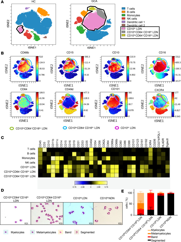
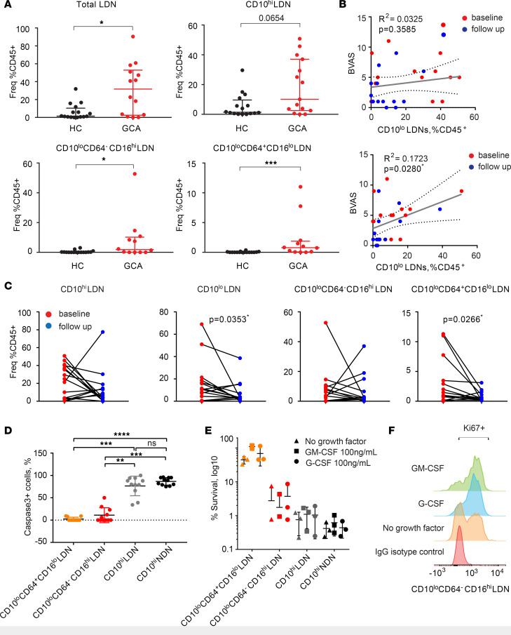
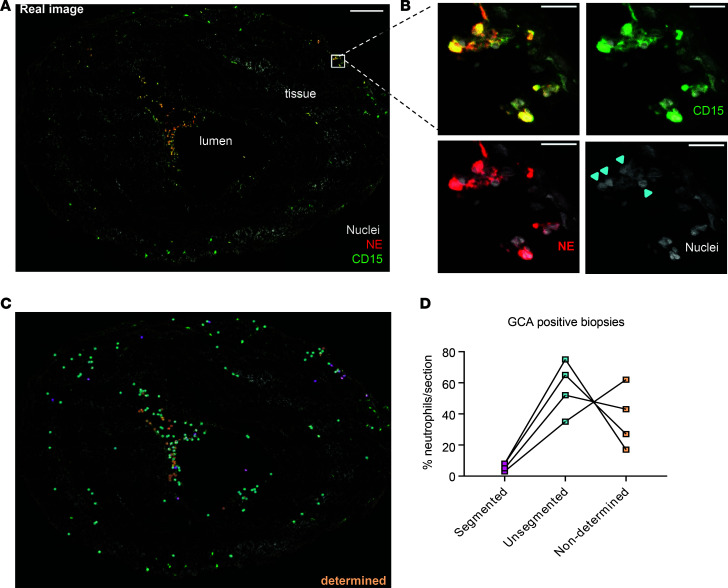
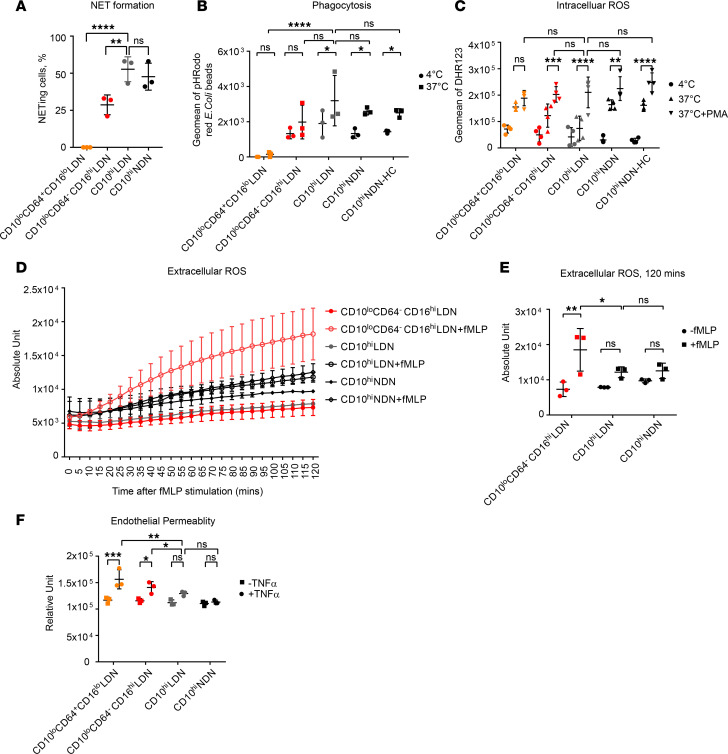
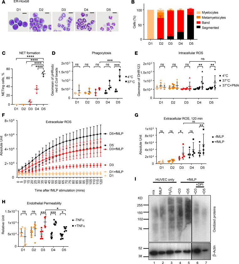
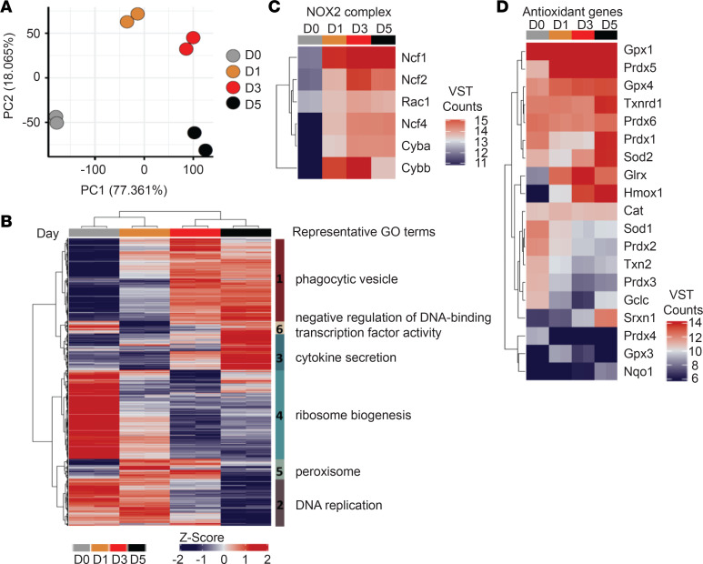
Giant cell arteritis (GCA) is a common form of primary systemic vasculitis in adults, with no reliable indicators of prognosis or treatment responses. We used single cell technologies to comprehensively map immune cell populations in the blood of patients with GCA and identified the CD66b+CD15+CD10lo/-CD64- band neutrophils and CD66bhiCD15+CD10lo/-CD64+/bright myelocytes/metamyelocytes to be unequivocally associated with both the clinical phenotype and response to treatment. Immature neutrophils were resistant to apoptosis, remained in the vasculature for a prolonged period of time, interacted with platelets, and extravasated into the tissue surrounding the temporal arteries of patients with GCA. We discovered that immature neutrophils generated high levels of extracellular reactive oxygen species, leading to enhanced protein oxidation and permeability of endothelial barrier in an in vitro coculture system. The same populations were also detected in other systemic vasculitides. These findings link functions of immature neutrophils to disease pathogenesis, establishing a clinical cellular signature of GCA and suggesting different therapeutic approaches in systemic vascular inflammation.

Keywords: Autoimmune diseases; Neutrophils; Vascular Biology; Vasculitis.


Conflict of interest statement

Conflict of interest: The authors have declared that no conflict of interest exists.

Figures

Figure 1
Figure 1. GCA patients are characterized by the presence of immature neutrophil populations in their blood.
(A) Samples were clustered using viSNE, and cell populations were identified by expression of the main canonical markers. Representative viSNE clustering plots for 1 healthy control (HC) and 1 GCA PBMC sample are shown. Circled in black are total low density neutrophils (LDNs). (B) Highlighted expression of key neutrophil surface markers on viSNE plots of PBMC from 1 representative GCA. (C) The expression levels of selected markers in each of the identified cell populations are shown in the expression heatmap from 1 representative GCA patient. (D) Wright-Giemsa staining of FACS purified 4 neutrophil populations. One representative from at least 3 independent experiments of 3 GCA patients is shown. (E) Quantification of neutrophil of different maturation stages in each LDN population showed that, while both CD10hi LDNs and NDNs were predominantly made of mature segmented neutrophils, myelocytes and metamylocytes contributed 80% of CD10loCD64+CD16lo LDNs, and 80% immature band neutrophils were found in CD10loCD64CD16hi LDNs. Data are presented as mean ± SD.
Figure 2
Figure 2. CD10lo LDNs with extended life span are clinically relevant to GCA.
(A) Neutrophil population frequency comparison between GCA and HC (GCA, n = 14; HC, n = 16). Unpaired nonparametric Mann-Whitney U test was used for statistical analysis. Data are presented with median ± IQR. (B) Correlation of CD10hi and CD10lo LDNs at baseline and follow-up after a single high-dose prednisolone treatment. Spearman’s correlation coefficient was calculated (baseline, n = 13; follow-up, n = 13). (C) Correlation of neutrophil subset frequency at baseline and follow-up. CD10loCD64+CD16lo and total CD10lo LDNs showed significant negative correlation. A nonparametric paired Wilcoxon test was used for statistical analysis to compare the difference of neutrophil populations between baseline and follow-up measurement within the same patients. (D) The apoptotic rate of immature CD10loCD64CD16hi and CD10loCD64+CD16lo LDNs was significantly reduced compared with mature CD10hi NDNs and LDNs after 24 hours in vitro culture as evidenced by caspase-3 staining (no growth factor in presence). Quantification of caspase-3–activated cells was performed by counting FITC+ out of the total DAPI+ cells. Three independent experiments were performed on FACS purified neutrophil populations from 3 GCA patients. The quantification was carried out by counting cells from 4–5 fields of a total of 100–200 cells of each population of each donor. Data are presented as mean ± SD. Nonparametric 1-way ANOVA Kruskal-Wallis test was used for statistical analysis. (E) CD10loCD64+CD16lo LDNs could survive up to 5 days in vitro, even without the presence of neutrophil growth factors such as G-CSF and GM-CSF. (F) CD10loCD64+CD16lo LDNs retained the proliferation capacity up to 3 days in culture measured by Ki67 expression, which is associated with cellular proliferation. Histograms from 1 representative GCA donor are shown. Three independent experiments were performed on FACS purified neutrophil populations from 3 GCA patients in E and F. *P ≤ 0.05, **P ≤ 0.01, ***P ≤ 0.001, ****P ≤ 0.0001.
Figure 3
Figure 3. Immature neutrophils extravasate into temporal artery walls of GCA patient biopsies.
(A) Confocal image of a temporal artery section of a GCA biopsy stained for neutrophil Elastase (NE, red), CD15 (green), and Hoechst (gray) for DNA revealed presence of neutrophils in both the lumen and tissue. Scale bar: 200 μm. (B) Zoomed-in regions in a gallery overview exemplify the morphology of both segmented and unsegmented nuclei within the specimen and emphasize the individual staining (from left to right and top to bottom: all combined, Hoechst + CD15, Hoechst + NE, Hoechst-only nuclei). Arrowheads indicate unsegmented neutrophil nuclei. Scale bar: 20 μm. (C) The point map of the whole section shows categorization of neutrophils based on the nuclear shape. (D) The respective quantification of neutrophils on 1 section from 4 different biopsies.
Figure 4
Figure 4. CD10lo LDNs are potent ROS producers but deficient in some innate immune functions.
(A) NET quantification by counting NET-forming cells stained positively with Cit-H3 and NE out of total DAPI+ cells. Three independent experiments were performed on FACS purified neutrophil populations from 3 GCA patients. (B) Phagocytosis was FACS quantified by the intake of pHrodo red E. coli bioparticles across the neutrophil populations. Mature but not immature neutrophils were capable of efficient phagocytosis. Three independent experiments were performed on FACS purified neutrophil populations from 3 GCA patients. (C) Intracellular ROS production was FACS quantified by green fluorescence converted from dihydrorhodamine 123 (DHR123) in the presence of ROS. Both mature and immature LDNs could generate ROS intracellularly, comparable with mature NDNs under PMA stimulation. Four independent experiments were performed on FACS purified neutrophil populations from 4 GCA and 4 HC donors. (D) Extracellular ROS was measured using OxyBURST Green H2HFF BSA. In the presence of 1 μM fMLP, immature CD10loCD64CD16hi LDNs showed an increased and sustained release of ROS in comparison with CD10hi LDNs and CD10hi NDNs up to 120 minutes. (E) Extracellular ROS production at 120 minute with or without fMLP treatment. (F) Vascular damage was quantified by a permeability assay of endothelial barrier in a neutrophil-endothelial coculture system. Both CD10loCD64CD16hi and CD10loCD64+CD16lo immature LDNs showed higher permeability compared with CD10hi NDNs, indicating their potential roles associated with vascular damage in vasculitis. Three independent experiments were performed on FACS purified neutrophil populations from 3 GCA patients in D, E, and F. Two-way ANOVA was used for statistical analysis in A, B, C, E, and F. Data are presented as mean ± SD. *P ≤ 0.05, **P ≤ 0.01, ***P ≤ 0.001, ****P ≤ 0.0001.
Figure 5
Figure 5. ER-Hoxb8–differentiated neutrophils faithfully recapitulate the phenotypes and functions of human neutrophil counterparts.
(A) Morphology of ER-Hoxb8–differentiated neutrophils from D1 to D5 in the presence of G-CSF. Scare bars: 10 μm. One representative from 3–5 independent experiments is shown. (B) Quantification of neutrophil of different maturation stages in each ER-Hoxb8–differentiated neutrophil defined by days after G-CSF treatment. Data from at least 3 independent experiments are shown. (C) NET quantification by counting NET-forming cells stained positively with Cit-H3 and NE out of total DAPI+ cells. (D) Only mature D5 Hoxb8 neutrophils were able to perform phagocytosis. (E) Immature D3 Hoxb8 neutrophils were competent intracellular ROS producer comparable with mature D5 counterpart. (F) Immature D3 Hoxb8 neutrophils were competent to generate extracellular ROS over a period of 120 minutes. Five-minute intervals were used to plot each time point. (G) Extracellular ROS production at 120 minutes was compared across ER-Hoxb8 neutrophils of different days of maturation. (H) D3 Hoxb8 neutrophils showed higher endothelial permeability compared with neutrophils of other maturation stages in neutrophil-endothelial coculture permeability system. Data from at least 3 independent experiments are shown from C to H. (I) Detection of oxidized protein in HUVECs with Hoxb8 neutrophil coculture. Oxidized proteins potentially caused by extracellular ROS generated by D3 and D5 Hoxb8 neutrophils in the presence of 1 μM fMLP were detected via carbonylated groups in HUVEC. HUVEC without any treatment (–fMLP) and with fMLP alone (+fMLP) were used as negative controls. HUVECs treated with 10 μM H2O2 were used as the positive control. One representative from 3 independent experiments is shown. Two-way ANOVA was performed for statistical analysis in C, D, E, G, and H. Data are presented as mean ± SD. *P ≤ 0.05, **P ≤ 0.01, ***P ≤ 0.001, ****P ≤ 0.0001.
Figure 6
Figure 6. Immature neutrophils display functional ROS biosynthesis system, but the antioxidant systems develop with maturation.
(A) The first 2 components from principle component analysis (PCA) of 11,412 differentially expressed (adjusted P < 0.01) are shown, which separate the cells by maturity. (B) Z-scores of differentially expressed genes (as in A). Hierarchical clustering of Euclidean distances reveals 6 distinct clusters, which differ in their pattern of expression across differentiation. Representative gene ontology (GO) terms are shown for each cluster. (C and D) Heatmaps of variance stabilized counts for differentially expressed genes comprising the NADPH oxidase 2 (NOX2) complex (C) and selected antioxidant genes (D).

References

    1. Luqmani R. Maintenance of clinical remission in ANCA-associated vasculitis. Nat Rev Rheumatol. 2013;9(2):127–132. doi: 10.1038/nrrheum.2012.188. - DOI - PubMed
    1. De Smit E, Palmer AJ, Hewitt AW. Projected worldwide disease burden from giant cell arteritis by 2050. J Rheumatol. 2015;42(1):119–125. doi: 10.3899/jrheum.140318. - DOI - PubMed
    1. Nakazawa D, Masuda S, Tomaru U, Ishizu A. Pathogenesis and therapeutic interventions for ANCA-associated vasculitis. Nat Rev Rheumatol. 2019;15(2):91–101. doi: 10.1038/s41584-018-0145-y. - DOI - PubMed
    1. Luqmani R, et al. The Role of Ultrasound Compared to Biopsy of Temporal Arteries in the Diagnosis and Treatment of Giant Cell Arteritis (TABUL): a diagnostic accuracy and cost-effectiveness study. Health Technol Assess. 2016;20(90):1–238. doi: 10.3310/hta20900. - DOI - PMC - PubMed
    1. de Boysson H, et al. 18F-fluorodeoxyglucose positron emission tomography and the risk of subsequent aortic complications in giant-cell arteritis: A multicenter cohort of 130 patients. Medicine (Baltimore) 2016;95(26):e3851. doi: 10.1097/MD.0000000000003851. - DOI - PMC - PubMed

Publication types

MeSH terms