Doxorubicin-induced p53 interferes with mitophagy in cardiac fibroblasts
- PMID: 32960902
- PMCID: PMC7508395
- DOI: 10.1371/journal.pone.0238856
Doxorubicin-induced p53 interferes with mitophagy in cardiac fibroblasts
Abstract
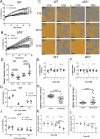
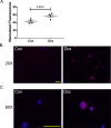
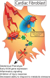
Anthracyclines are the critical component in a majority of pediatric chemotherapy regimens due to their broad anticancer efficacy. Unfortunately, the vast majority of long-term childhood cancer survivors will develop a chronic health condition caused by their successful treatments and severe cardiac disease is a common life-threatening outcome that is unequivocally linked to previous anthracycline exposure. The intricacies of how anthracyclines such as doxorubicin, damage the heart and initiate a disease process that progresses over multiple decades is not fully understood. One area left largely unstudied is the role of the cardiac fibroblast, a key cell type in cardiac maturation and injury response. In this study, we demonstrate the effect of doxorubicin on cardiac fibroblast function in the presence and absence of the critical DNA damage response protein p53. In wildtype cardiac fibroblasts, doxorubicin-induced damage correlated with decreased proliferation and migration, cell cycle arrest, and a dilated cardiomyopathy gene expression profile. Interestingly, these doxorubicin-induced changes were completely or partially restored in p53-/- cardiac fibroblasts. Moreover, in wildtype cardiac fibroblasts, doxorubicin produced DNA damage and mitochondrial dysfunction, both of which are well-characterized cell stress responses induced by cytotoxic chemotherapy and varied forms of heart injury. A 3-fold increase in p53 (p = 0.004) prevented the completion of mitophagy (p = 0.032) through sequestration of Parkin. Interactions between p53 and Parkin increased in doxorubicin-treated cardiac fibroblasts (p = 0.0003). Finally, Parkin was unable to localize to the mitochondria in wildtype cardiac fibroblasts, but mitochondrial localization was restored in p53-/- cardiac fibroblasts. These findings strongly suggest that cardiac fibroblasts are an important myocardial cell type that merits further study in the context of doxorubicin treatment. A more robust knowledge of the role cardiac fibroblasts play in the development of doxorubicin-induced cardiotoxicity will lead to novel clinical strategies that will improve the quality of life of cancer survivors.
Conflict of interest statement
NO authors have competing interest.
Figures


Similar articles
-
Cardiomyocyte specific ablation of p53 is not sufficient to block doxorubicin induced cardiac fibrosis and associated cytoskeletal changes.PLoS One. 2011;6(7):e22801. doi: 10.1371/journal.pone.0022801. Epub 2011 Jul 28. PLoS One. 2011. PMID: 21829519 Free PMC article.
-
SESN2 protects against doxorubicin-induced cardiomyopathy via rescuing mitophagy and improving mitochondrial function.J Mol Cell Cardiol. 2019 Aug;133:125-137. doi: 10.1016/j.yjmcc.2019.06.005. Epub 2019 Jun 12. J Mol Cell Cardiol. 2019. PMID: 31199952
-
Inhibition of AMP-activated protein kinase α (AMPKα) by doxorubicin accentuates genotoxic stress and cell death in mouse embryonic fibroblasts and cardiomyocytes: role of p53 and SIRT1.J Biol Chem. 2012 Mar 9;287(11):8001-12. doi: 10.1074/jbc.M111.315812. Epub 2012 Jan 20. J Biol Chem. 2012. PMID: 22267730 Free PMC article.
-
Role of mitochondria in doxorubicin-mediated cardiotoxicity: From molecular mechanisms to therapeutic strategies.Cell Stress Chaperones. 2024 Apr;29(2):349-357. doi: 10.1016/j.cstres.2024.03.003. Epub 2024 Mar 12. Cell Stress Chaperones. 2024. Retraction in: Cell Stress Chaperones. 2024 Oct;29(5):641. doi: 10.1016/j.cstres.2024.08.003. PMID: 38485043 Free PMC article. Retracted. Review.
-
Autophagy and mitophagy in the context of doxorubicin-induced cardiotoxicity.Oncotarget. 2017 Jul 11;8(28):46663-46680. doi: 10.18632/oncotarget.16944. Oncotarget. 2017. PMID: 28445146 Free PMC article. Review.
Cited by
-
Moxibustion alleviates chronic heart failure by regulating mitochondrial dynamics and inhibiting autophagy.Exp Ther Med. 2022 May;23(5):359. doi: 10.3892/etm.2022.11286. Epub 2022 Mar 30. Exp Ther Med. 2022. PMID: 35493422 Free PMC article.
-
Doxorubicin-induced modulation of TGF-β signaling cascade in mouse fibroblasts: insights into cardiotoxicity mechanisms.Sci Rep. 2023 Nov 2;13(1):18944. doi: 10.1038/s41598-023-46216-7. Sci Rep. 2023. PMID: 37919370 Free PMC article.
-
A safety screening platform for individualized cardiotoxicity assessment.iScience. 2024 Feb 6;27(3):109139. doi: 10.1016/j.isci.2024.109139. eCollection 2024 Mar 15. iScience. 2024. PMID: 38384853 Free PMC article.
-
Live cell, image-based high-throughput screen to quantitate p53 stabilization and viability in human papillomavirus positive cancer cells.Virology. 2021 Aug;560:96-109. doi: 10.1016/j.virol.2021.05.006. Epub 2021 May 22. Virology. 2021. PMID: 34051479 Free PMC article.
-
Overcoming doxorubicin resistance in triple-negative breast cancer using the class I-targeting HDAC inhibitor bocodepsin/OKI-179 to promote apoptosis.Breast Cancer Res. 2024 Mar 1;26(1):35. doi: 10.1186/s13058-024-01799-5. Breast Cancer Res. 2024. PMID: 38429789 Free PMC article.
References
-
- Howlader N NA, Krapcho M, Garshell J, Miller D, Altekruse SF, Kosary CL, et al. SEER Cancer Statistics Review, 1975–2012. Bethesda, MD: National Cancer Institute, 2015.
-
- Armstrong GT, Kawashima T, Leisenring W, Stratton K, Stovall M, Hudson MM, et al. Aging and risk of severe, disabling, life-threatening, and fatal events in the childhood cancer survivor study. Journal of clinical oncology: official journal of the American Society of Clinical Oncology. 2014;32(12):1218–27. Epub 2014/03/19. 10.1200/jco.2013.51.1055 - DOI - PMC - PubMed
Publication types
MeSH terms
Substances
Grants and funding
LinkOut - more resources
Full Text Sources
Research Materials
Miscellaneous

