Review
. 2020 Nov-Dec;22(10):515-524.
doi: 10.1016/j.micinf.2020.09.004. Epub 2020 Sep 19.

Vaccines for COVID-19: perspectives from nucleic acid vaccines to BCG as delivery vector system

Affiliations
Review

Vaccines for COVID-19: perspectives from nucleic acid vaccines to BCG as delivery vector system

Nina Marí G P de Queiroz et al. Microbes Infect. 2020 Nov-Dec.

Abstract

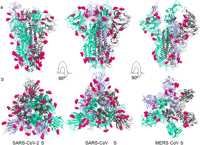
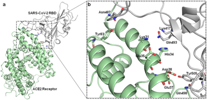
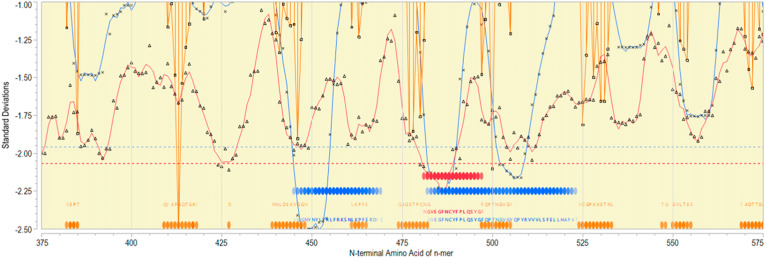
This article discusses standard and new disruptive strategies in the race to develop an anti-COVID-19 vaccine. We also included new bioinformatic data from our group mapping immunodominant epitopes and structural analysis of the spike protein. Another innovative approach reviewed here is the use of BCG vaccine as priming strategy and/or delivery system expressing SARS-CoV-2 antigens.

Keywords: BCG; COVID-19; RBD domain; SARS-CoV-2; Vaccines.


Conflict of interest statement

Declaration of competing interest EJH is an employee and equity holder in ioGenetics LLC. The funders had no role in the design of the study; in the collection, analyses, or interpretation of data; in the writing of the manuscript, or in the decision to publish the results.

Figures

Fig. 1
Fig. 1
Schematic organization of SARS-Cov-2 genome. The ORF1a and ORF1b comprise about 67% of the genome encoding 16 nonstructural proteins (nsps). In addition to nsps, the genome encodes the four major structural proteins: spike (S), membrane (M), nucleocapsid (N) and envelope (E).
Fig. 2
Fig. 2
Structural comparison between the SARS-CoV-2 S (PDB ID: 6VSB), SARS-CoV S (PDB ID: 5X58) and MERS-CoV S (PDB ID: 5X59) proteins. The individual protomer of S protein trimer domain is represented in a different color: violet, clear green and gray, respectively. a) Side and b) top conformation views of the prefusion structure of the S glycoproteins with N-linked glycans rendered as hot pink spheres.
Fig. 3
Fig. 3
Structure of SARS-CoV-2 RBD in fusion conformation and interaction with the angiotensin-converting enzyme 2 (ACE2) (PDB ID: 6M0J). Proteins are individually colored in gray (SARS-CoV-2 RBD) and green (ACE2 Receptor). Sticks representation with colors by elements show the amino acid residues Tyr83, Lys31, Glu35, His34, Asp38, Glu37 in ACE2 receptor and Asn487, Lys417, Gln493, Tyr505 and Gln498, residues in SARS-CoV-2 RBD.
Fig. 4
Fig. 4
Epitope mapping of RBD region of Spike protein. X axis indicates sequential peptides with single amino acid displacement. Y axis indicates predicted binding affinity in SD units for the protein. The red line shows the permuted average predicted MHC-IA and B (37 alleles) binding affinity by index position of sequential 9-mer peptides with single amino acid displacement. The blue line shows the permuted average predicted MHC-II DRB allele (16 most common human alleles) binding affinity by index position of sequential 15-mer peptides. Both are plotted in standard deviation units, shown on the Y axis. Orange lines show the predicted probability of B-cell receptor binding for an amino acid centered in each sequential 9-mer peptide. Low numbers for MHC data represent high binding affinity, whereas low numbers equate to high B cell receptor contact probability. Ribbons (red: MHC-I, blue: MHC-II) indicate the 10% highest predicted MHC affinity binding. Orange ribbons indicate the top 25% predicted probability B-cell binding. Horizontal dotted lines demarcate the top 5% of binding affinity for the protein (red MHC I, blue MHC II).

References

    1. WHO Summary of MERS cases with onset of illness from 1 September 2012. https://www.who.int/emergencies/mers-cov/en/ Available at:
    1. Xu X., Chen P., Wang J., Feng J., Zhou H., Li X. Evolution of the novel coronavirus from the ongoing Wuhan outbreak and modeling of its spike protein for risk of human transmission. Sci China Life Sci. 2020;63:457–460. - PMC - PubMed
    1. Raj V.S., Mou H., Smits S.L., Dekkers D.H., Muller M.A., Dijkman R. Dipeptidyl peptidase 4 is a functional receptor for the emerging human coronavirus-EMC. Nature. 2013;495:251–254. - PMC - PubMed
    1. Snijder E.J., van der Meer Y., Zevenhoven-Dobbe J., Onderwater J.J., van der Meulen J., Koerten H.K. Ultrastructure and origin of membrane vesicles associated with the severe acute respiratory syndrome coronavirus replication complex. J Virol. 2006;80:5927–5940. - PMC - PubMed
    1. Hussain S., Pan J., Chen Y., Yang Y., Xu J., Peng Y. Identification of novel subgenomic RNAs and noncanonical transcription initiation signals of severe acute respiratory syndrome coronavirus. J Virol. 2005;79:5288–5295. - PMC - PubMed

Publication types

MeSH terms