. 2020 Nov:175:107314.
doi: 10.1016/j.nlm.2020.107314. Epub 2020 Sep 20.

Persistent changes in extracellular lactate dynamics following synaptic potentiation

Affiliations

Persistent changes in extracellular lactate dynamics following synaptic potentiation

D Bingul et al. Neurobiol Learn Mem. 2020 Nov.

Abstract

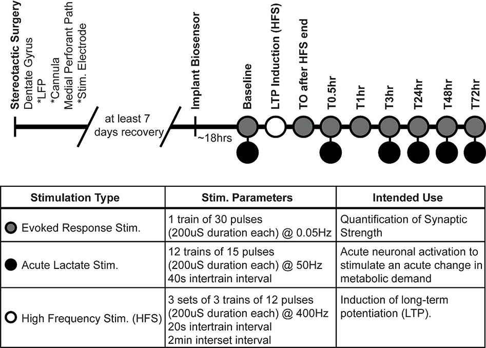
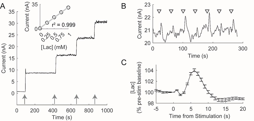
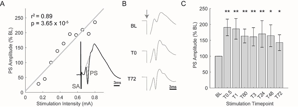
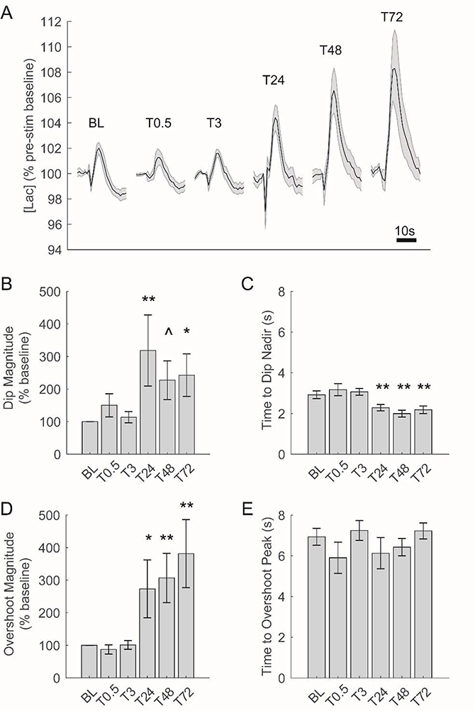
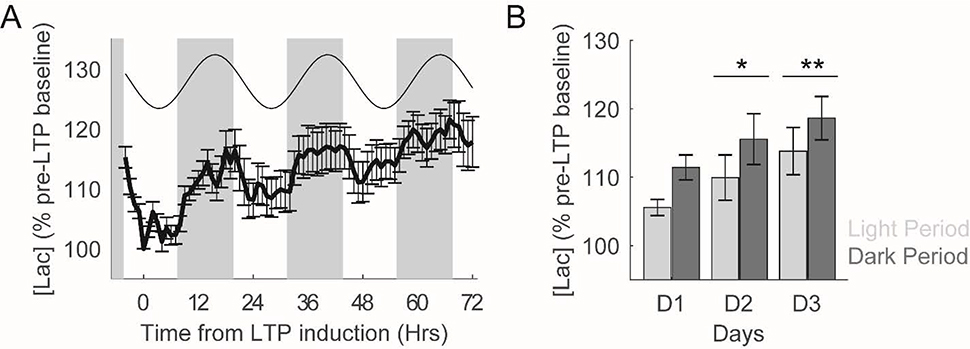
A diverse array of neurometabolic coupling mechanisms exist within the brain to ensure that sufficient metabolite availability is present to meet both acute and chronic energetic demands. Excitatory synaptic activity, which produces the majority of the brain's energetic demands, triggers a rapid metabolic response including a characteristic shift towards aerobic glycolysis. Herein, astrocytically derived lactate appears to serve as an important metabolite to meet the extensive metabolic needs of activated neurons. Despite a wealth of literature characterizing lactate's role in mediating these acute metabolic needs, the extent to which lactate supports chronic energetic demands of neurons remains unclear. We hypothesized that synaptic potentiation, a ubiquitous brain phenomenon that can produce chronic alterations in synaptic activity, could necessitate persistent alterations in brain energetics. In freely-behaving rats, we induced long-term potentiation (LTP) of synapses within the dentate gyrus through high-frequency electrical stimulation (HFS) of the medial perforant pathway. Before, during, and after LTP induction, we continuously recorded extracellular lactate concentrations within the dentate gyrus to assess how changes in synaptic strength alter local glycolytic activity. Synaptic potentiation 1) altered the acute response of extracellular lactate to transient neuronal activation as evident by a larger initial dip and subsequent overshoot and 2) chronically increased local lactate availability. Although synapses were potentiated immediately following HFS, observed changes in lactate dynamics were only evident beginning ~24 h later. Once observed, however, both synaptic potentiation and altered lactate dynamics persisted for the duration of the experiment (~72 h). Persistent alterations in synaptic strength, therefore, appear to be associated with metabolic plasticity in the form of persistent augmentation of glycolytic activity.

Keywords: Glycolysis; Hippocampus; Lactate; Long-term potentiation; Synaptic plasticity.


Figures

Figure 1.
Figure 1.
Experimental timeline and stimulation parameters. Within the dentate gyrus, an implanted local field potential and lactate-sensitive biosensor were used to record evoked responses and extracellular [lac], respectively. Electrical stimulations of the medial perforant path were used to quantify synaptic strength, produce acute neuronal activation and associated changes in metabolic demand, and produce lasting synaptic potentiation within the dentate gyrus. At time points with two stimulation types, evoked response stimulation always preceded acute lactate stimulations.
Figure 2.
Figure 2.
Amperometric biosensors are sensitive to in vitro and in vivo changes in lactate concentration ([lac]). A) In vitro calibration of a lactate sensitive biosensor. Arrows depict the addition of lactate (0.25mM). Lactate biosensors rapidly (~1s) produce an increase in current in response to changes in [Lac] that is linear across the expected range of [Lac] in vivo (see inset). B) Baseline electrical stimulations of the perforant path in an individual produce reliable changes in extracellular [lac]. Triangles depict each stimulation wherein 15, 200uS pulses were delivered at 50Hz. C) Average change in [Lac] observed during baseline stimulation in an individual rat. Following stimulation, extracellular [Lac] initially decreases followed by an overshoot of pre-stimulation levels.
Figure 3.
Figure 3.
High frequency stimulation (HFS) significantly potentiates the population spike of the evoked response for at least 72hrs. A) Typical input/output curve used to determine the baseline (BL) intensity of perforant path stimulation. BL intensity was chosen to produce an evoked response that contained a population spike equal to ~40% of the maximal population spike that could be elicited. Inset depicts a typical evoked response (SA: stimulation artifact, PS: population spike). B) Average evoked response observed during three separate stimulation periods in an individual rat (stimulation artifacts have been truncated to better visualize the evoked response). Grey arrow depicts time of stimulation. HFS was administered between BL and T0. C) Average population spike observed during each stimulation period (N = 9 rats; **p<0.01, *p<0.05 as compared to BL).
Figure 4.
Figure 4.
Changes in [lac] in response to acute electrical stimulation are augmented beginning 24hrs after induction of LTP. A) Average change in [lac] in response to acute electrical stimulation across all rats at each stimulation time point. Each tracing depicts 25s of [lac] (including 5s prior to stimulation). BCDE) Changes in [lac] following acute electrical stimulation before and after LTP induction. 24hrs after LTP induction, the initial decrease in [lac] following stimulation is both significantly larger (B) and faster (C) while the magnitude of ensuing [lac] overshoot is also significantly increased (D). **p<0.01, *p<0.05, and ^p<0.10 as compared to baseline (BL).
Figure 5.
Figure 5.
LTP induction produces persistent increases in extracellular [lac]. A) Extracellular [lac] across 72hrs post-LTP induction. In addition to exhibiting a clear circadian rhythmicity, [lac] are consistently increased across the days following LTP induction. Grey boxes depict lights off. Best fitting cosine function to model circadian rhythmicity is presented above [lac] tracing. B) Average extracellular [lac] during the light and dark periods during the three days of data depicted in A. [Lac] are significantly higher during the dark period as compared to the light period. Moreover, relative to the day of LTP induction (D1), [lac] are significantly higher during each of the next two days. **p<0.01 and *p<0.05.

References

    1. Abraham WC, Williams JM (2008) LTP maintenance and its protein synthesis-dependence. Neurobiology of Learning and Memory 89:260–268. - PubMed
    1. Alle H, Roth A, Geiger JR (2009) Energy-efficient action potentials in hippocampal mossy fibers. Science 325:1405–1408. - PubMed
    1. Attwell D, Gibb A (2005) Neuroenergetics and the kinetic design of excitatory synapses. Nat Rev Neurosci 6:841–849. - PubMed
    1. Aubert A, Costalat R (2005) Interaction between Astrocytes and Neurons Studied using a Mathematical Model of Compartmentalized Energy Metabolism: Journal of Cerebral Blood Flow & Metabolism 25:1476–1490. - PubMed
    1. Aubert A, Costalat R, Magistretti PJ, Pellerin L (2005) Brain lactate kinetics: Modeling evidence for neuronal lactate uptake upon activation. PNAS 102:16448–16453. - PMC - PubMed

Publication types