Review
. 2021 Feb:68:28-36.
doi: 10.1016/j.ceb.2020.08.014. Epub 2020 Sep 19.

Mitochondrial dynamics: Shaping and remodeling an organelle network

Affiliations
Review

Mitochondrial dynamics: Shaping and remodeling an organelle network

Adam R Fenton et al. Curr Opin Cell Biol. 2021 Feb.

Abstract

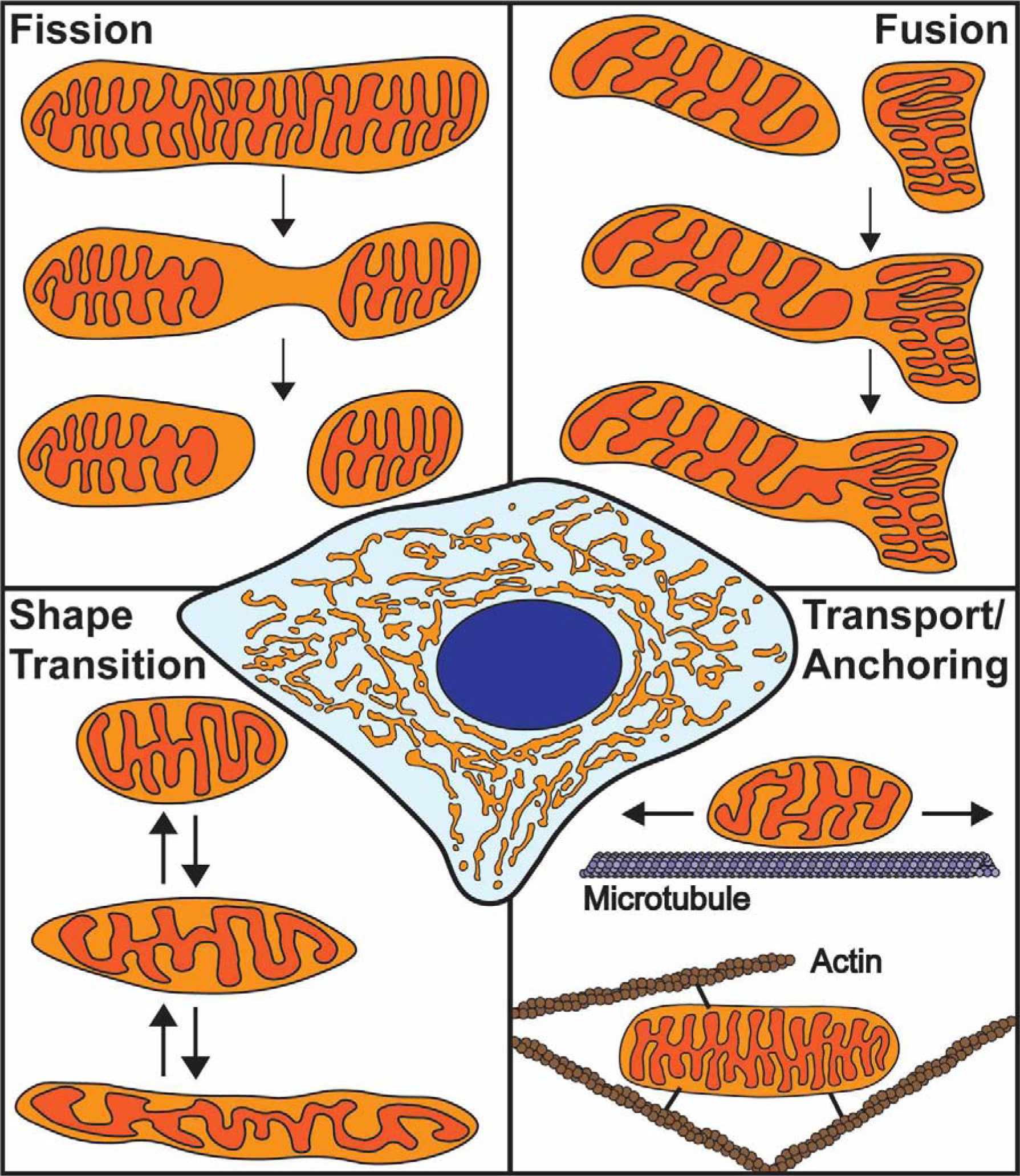
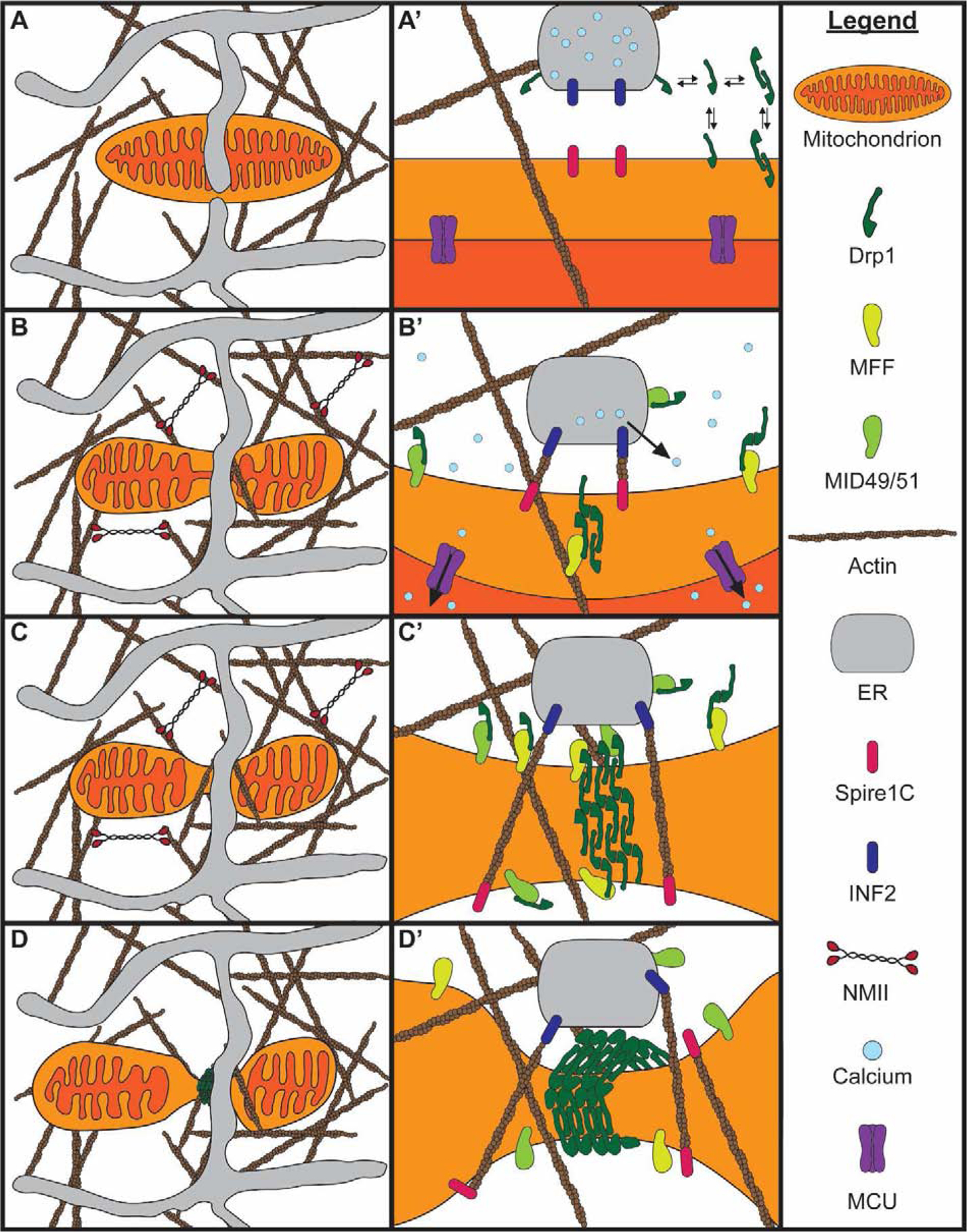
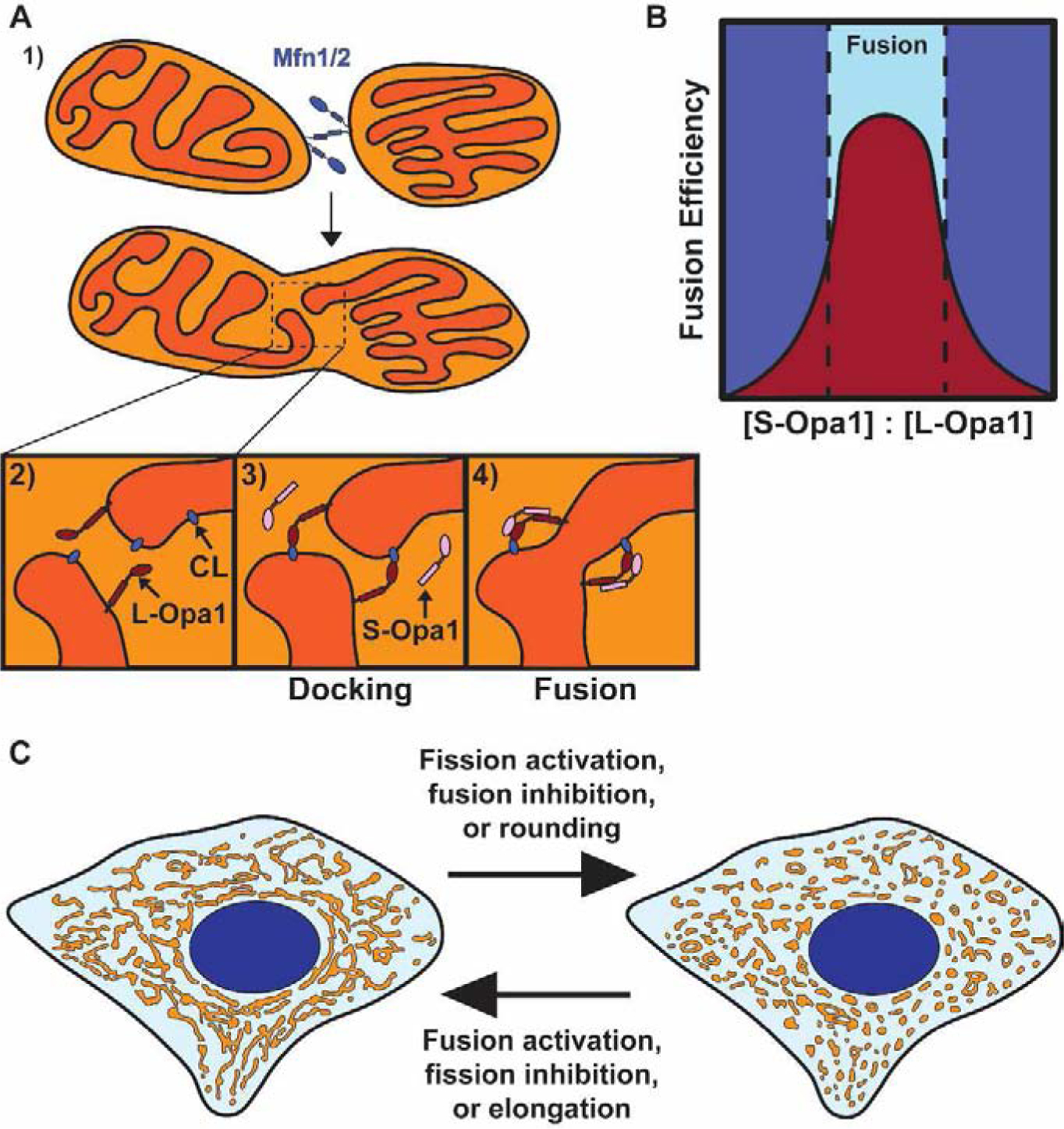
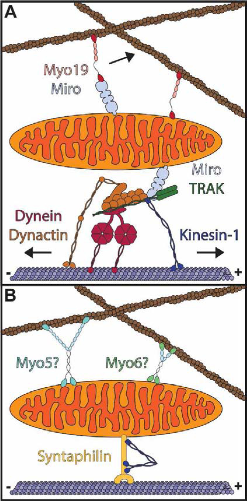
Mitochondria form networks that continually remodel and adapt to carry out their cellular function. The mitochondrial network is remodeled through changes in mitochondrial morphology, number, and distribution within the cell. Mitochondrial dynamics depend directly on fission, fusion, shape transition, and transport or tethering along the cytoskeleton. Over the past several years, many of the mechanisms underlying these processes have been uncovered. It has become clear that each process is precisely and contextually regulated within the cell. Here, we discuss the mechanisms regulating each aspect of mitochondrial dynamics, which together shape the network as a whole.

Keywords: Cytoskeleton; Fission; Fusion; Mitochondria; Morphology; Transport.


Conflict of interest statement

Conflict of interest statement Nothing declared.

Figures

Figure 1.
Figure 1.. Overview of mitochondrial dynamics.
Mitochondria form a complex, interconnected network within the cell (center). The morphology of this network is determined by fission, fusion, mitochondrial shape transition, and positioning along the cytoskeleton. Fission begins with IMM division followed by OMM scission. Fusion involves merging of two outer membranes followed by joining of the inner membranes. Mitochondrial shape transition is a process independent of fission/fusion that controls the transition between rounded and elongated mitochondrial morphologies. Mitochondrial positioning involves transport and tethering along the microtubule and actin cytoskeletons.
Figure 2.
Figure 2.. Mechanism of mitochondrial fission.
A) A mitochondrion is embedded in the interstitial actin network and closely associated with the ER. A closer view of the mitochondria-ER contact is shown in A’. Drp1 dynamically associates with the cytosol, mitochondria, and ER prior to fission. B) Peripheral NMII pulls on actin filaments to deform the mitochondrial membrane. Increased cytosolic calcium induces actin polymerization at mitochondria-ER contacts by INF2 on the ER and Spire1C on mitochondria. Mff and Mid49/51 begin recruiting Drp1 to the mitochondria-ER contact. Calcium is released from the ER and enters the mitochondria through the MCU, causing IMM constriction. C) Elevated mitochondrial matrix calcium causes IMM division prior to OMM division. Mff and Mid49/51 continue recruiting Drp1 to the mitochondria-ER contact, with some Drp1 coming from the ER. Drp1 oligomerizes along the constricted OMM. D) The Drp1 oligomer fully assembles around the OMM. Drp1 GTP hydrolysis dissociates Mid49/51, constricting the Drp1 ring. The Drp1 ring constricts the OMM and completes the process of fission.
Figure 3.
Figure 3.. Mitochondrial fusion and shape transition.
A) Proposed model of mitochondrial fusion. 1) Mitochondrial fusion begins with Mfn1/2-mediated tethering of two mitochondrial outer membranes. 2) The inner membranes are positioned for fusion upon outer membrane fusion. 3) Interactions between L-Opa1 and cardiolipin (CL) dock the inner membranes, bringing them closer together. 4) S-Opa1 functions with L-Opa1 and cardiolipin to promote efficient inner membrane fusion. B) Fusion efficiency at different S-Opa1:L-Opa1 ratios. Fusion efficiency peaks at an equimolar ratio of S-Opa1 to L-Opa1, with higher and lower ratios inhibiting fusion. C) Schematic of a connected mitochondrial network (left) and fragmented mitochondrial network (right). The transition between these networks can occur through direct regulation of fission, fusion, or mitochondrial shape transition.
Figure 4.
Figure 4.. Mitochondrial transport and anchoring on the cytoskeleton.
A) TRAK and Miro proteins serve as adaptors for microtubule-based mitochondrial transport (below). Kinesin-1 drives transport to the microtubule plus-end while transport to the microtubule minus end is mediated by dynein/dynactin. Myo19 associates with Miro proteins and directly with the mitochondrial outer membrane to drive mitochondrial transport along the actin cytoskeleton. B) Mitochondria are anchored to the actin and microtubule cytoskeletons. Syntaphilin anchors mitochondria to microtubules while myosin V (Myo5), myosin VI (Myo6), or another tether may anchor mitochondria to actin.

References

    1. Ji W, Hatch AL, Merrill RA, Strack S & Higgs HN Actin filaments target the oligomeric maturation of the dynamin GTPase Drp1 to mitochondrial fission sites. eLife 4, e11553 (2015). - PMC - PubMed
    1. Ji W-K et al. Receptor-mediated Drp1 oligomerization on endoplasmic reticulum. J. Cell Biol 216, 4123–4139 (2017). - PMC - PubMed
    1. Otera H et al. Mff is an essential factor for mitochondrial recruitment of Drp1 during mitochondrial fission in mammalian cells. J. Cell Biol 191, 1141–1158 (2010). - PMC - PubMed
    1. Yu R et al. MIEF1/2 function as adaptors to recruit Drp1 to mitochondria and regulate the association of Drp1 with Mff. Sci. Rep 7, 880 (2017). - PMC - PubMed
    1. Palmer CS et al. Adaptor Proteins MiD49 and MiD51 Can Act Independently of Mff and Fis1 in Drp1 Recruitment and Are Specific for Mitochondrial Fission. J. Biol. Chem 288, 27584–27593 (2013). - PMC - PubMed

Publication types

Substances