Review
. 2020 Sep 19;21(18):6881.
doi: 10.3390/ijms21186881.

Metabolism of Glycosphingolipids and Their Role in the Pathophysiology of Lysosomal Storage Disorders

Affiliations
Review

Metabolism of Glycosphingolipids and Their Role in the Pathophysiology of Lysosomal Storage Disorders

Alex E Ryckman et al. Int J Mol Sci. .

Abstract

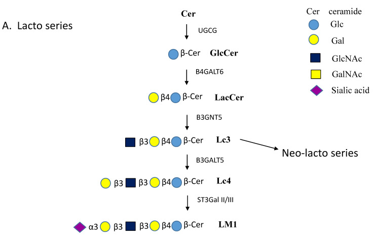
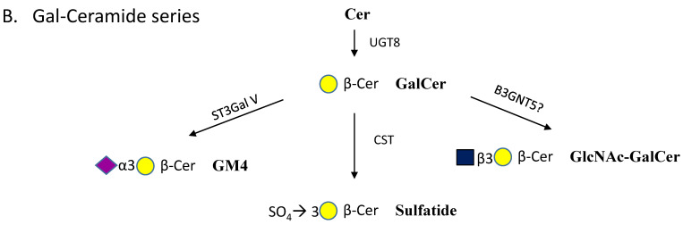
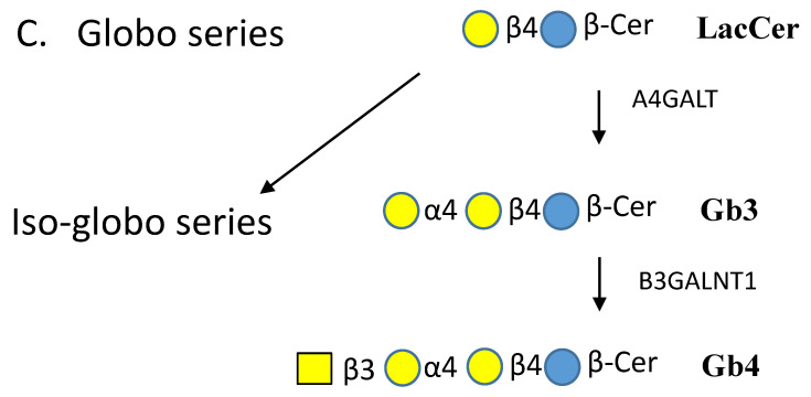
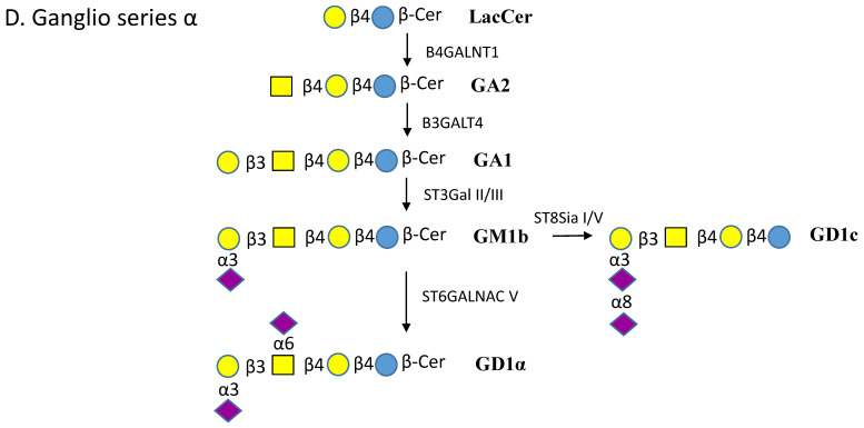
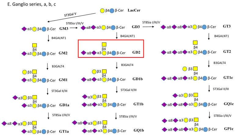
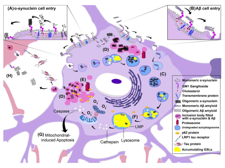
Glycosphingolipids (GSLs) are a specialized class of membrane lipids composed of a ceramide backbone and a carbohydrate-rich head group. GSLs populate lipid rafts of the cell membrane of eukaryotic cells, and serve important cellular functions including control of cell-cell signaling, signal transduction and cell recognition. Of the hundreds of unique GSL structures, anionic gangliosides are the most heavily implicated in the pathogenesis of lysosomal storage diseases (LSDs) such as Tay-Sachs and Sandhoff disease. Each LSD is characterized by the accumulation of GSLs in the lysosomes of neurons, which negatively interact with other intracellular molecules to culminate in cell death. In this review, we summarize the biosynthesis and degradation pathways of GSLs, discuss how aberrant GSL metabolism contributes to key features of LSD pathophysiology, draw parallels between LSDs and neurodegenerative proteinopathies such as Alzheimer's and Parkinson's disease and lastly, discuss possible therapies for patients.

Keywords: biosynthesis; degradation of glycoconjugates; glycosphingolipids; glycosyl hydrolases; lysosomal storage diseases; pathophysiology.


Conflict of interest statement

The authors declare no conflict of interest.

Figures

Figure 1
Figure 1
Pathways of glycosphingolipid (GSL) biosynthesis. (A). Lacto series GSLs. Lactosylceramide (LacCer) is the common intermediate for lacto, globo and ganglio series GSLs. The lacto series GSLs can have numerous different extensions and can carry glycan epitopes and antigens. Addition of GlcNAc to LacCer produces the precursor to all neo-lacto series GSLs, known as lactotriaosylceramide (Lc3). Lc3 is extended by the substitution of GlcNAc by a Galβ1-4 residue. The names of enzymes involved are indicated near the arrows. (B). Gal-ceramide series GSLs. Gal is added to ceramide by Cer-β-Gal-transferase UGT8 and reaction product GalCer can then undergo at least three different substitutions. (C). Globo series GSLs. The globosides are first produced by substitution of the Gal residue of LacCer with a Galα1-4 residue to yield globotriaosylceramide (Gb3). Addition of GalNAc in β1-3 linkage to Gb3 forms globotetraosylceramide (Gb4). The iso-globo series contains Galα1-3 instead of Galα1-4. (D). Ganglioseries-α GSLs. LacCer is the precursor for neutral GSLs, GA2 and GA1, and for all gangliosides. The addition of sialic acid in α2-6 linkage to GalNAc forms the α series of compounds. Here we show the synthesis of GD1α from GM1b. (E). Ganglio series a, b and c GSLs. The GM3 synthase ST3GAL V adds a sialic acid in α2-3 linkage to the Gal residue of LacCer to form GM3. The series that carries one sialic acid on Galβ1-4 is named ganglio a-series. Gangliosides having sialylα2-8sialylα2-3 substitutions are name ganglio b-series. The c-series have sialylα2-8sialylα2-8sialylα2-3 substitutions.
Figure 1
Figure 1
Pathways of glycosphingolipid (GSL) biosynthesis. (A). Lacto series GSLs. Lactosylceramide (LacCer) is the common intermediate for lacto, globo and ganglio series GSLs. The lacto series GSLs can have numerous different extensions and can carry glycan epitopes and antigens. Addition of GlcNAc to LacCer produces the precursor to all neo-lacto series GSLs, known as lactotriaosylceramide (Lc3). Lc3 is extended by the substitution of GlcNAc by a Galβ1-4 residue. The names of enzymes involved are indicated near the arrows. (B). Gal-ceramide series GSLs. Gal is added to ceramide by Cer-β-Gal-transferase UGT8 and reaction product GalCer can then undergo at least three different substitutions. (C). Globo series GSLs. The globosides are first produced by substitution of the Gal residue of LacCer with a Galα1-4 residue to yield globotriaosylceramide (Gb3). Addition of GalNAc in β1-3 linkage to Gb3 forms globotetraosylceramide (Gb4). The iso-globo series contains Galα1-3 instead of Galα1-4. (D). Ganglioseries-α GSLs. LacCer is the precursor for neutral GSLs, GA2 and GA1, and for all gangliosides. The addition of sialic acid in α2-6 linkage to GalNAc forms the α series of compounds. Here we show the synthesis of GD1α from GM1b. (E). Ganglio series a, b and c GSLs. The GM3 synthase ST3GAL V adds a sialic acid in α2-3 linkage to the Gal residue of LacCer to form GM3. The series that carries one sialic acid on Galβ1-4 is named ganglio a-series. Gangliosides having sialylα2-8sialylα2-3 substitutions are name ganglio b-series. The c-series have sialylα2-8sialylα2-8sialylα2-3 substitutions.
Figure 2
Figure 2
Schematic model of proposed pathophysiological mechanisms of lysosomal storage disorders (LSDs). The model is of the most affected cell type of LSDs, the neuron, and illustrates the events, which may indirectly or directly lead to cell death. A legend is presented on the right, which defines the symbols used in the figure. Several events are occurring simultaneously inside the diseased neuron: (A) α-synuclein cell entry, (B) Aβ cell entry, (C) accumulation of undegraded autophagosomes, (D) accumulation of cytoplasmic inclusion bodies filled with α-synuclein, Aβ or the Tau protein, (E) inhibition of the proteasome, (F) lysosomal membrane permeability changes, (G) mitochondrial-induced apoptosis (H) and cell–cell spreading of pathology. (A) α-synuclein enters the cell via GM1-receptor mediated endocytosis or by creating a pore in the membrane leading to the formation of inclusion bodies (seen in pink) that inhibit the proteasome and reduce degradation of materials in the cell. (B) Aβ amyloid enters the cell via the same method: GM1-mediated endocytosis. (C) Autophagosomes fill with undegraded material such as the accumulating proteins α-synuclein, Aβ and Tau. (F) Due to defective lysosomal hydrolases, GSLs accumulate in the lysosome and promote lysosomal membrane permeability (LMP). (G) Eventually the lysosome expels its contents including cathepsins. This induces mitochondrial release of caspases, which in turn cause apoptosis. Additionally, monomeric α-synuclein is capable of inhibiting mitochondrial fusion. As a result, the dying mitochondria degrade in the cytoplasm, releasing copious amounts of reactive oxygen species inducing cell death. (H) As the cell fills with undegraded autophagosomes, and harmful inclusion bodies, the cell activates secretory pathways that lead to the exocytosis of intracellular material to reduce the load, followed by endocytosis in adjacent cells. This provides a route for pathology to spread to adjacent cells. Harmful aggregates such as from Tau, α-synuclein and Aβ can be endocytosed into the adjacent cell. Specifically, Tau can interact with the receptor LRP1 to allow cell entry.

References

    1. Russo D., Capolupo L., Loomba J.S., Sticco L., D’Angelo G. Glycosphingolipid metabolism in cell fate specification. J. Cell Sci. 2018;131:jcs219204. doi: 10.1242/jcs.219204. - DOI - PubMed
    1. Ledeen R., Wu G. Gangliosides of the nervous system. Methods Mol. Biol. 2018;1804:19–55. - PubMed
    1. Gault C.R., Obeid L.M., Hannun Y.A. An overview of sphingolipid metabolism: From synthesis to breakdown. Adv. Exp. Med. Biol. 2010;688:1–23. - PMC - PubMed
    1. Honke K. Biosynthesis and biological function of sulfoglycolipids. Proc. Jpn. Acad. Ser. B Phys. Biol. Sci. 2013;89:129–138. doi: 10.2183/pjab.89.129. - DOI - PMC - PubMed
    1. Sonnino S., Chiricozzi E., Grassi S., Mauri L., Prioni S., Prinetti A. Gangliosides in membrane organization. Prog. Mol. Biol. Transl. Sci. 2018;156:83–120. - PubMed