. 2020 Sep 19;21(18):6890.
doi: 10.3390/ijms21186890.

Cell-Penetrable Peptide-Conjugated FADD Induces Apoptosis and Regulates Inflammatory Signaling in Cancer Cells

Affiliations

Cell-Penetrable Peptide-Conjugated FADD Induces Apoptosis and Regulates Inflammatory Signaling in Cancer Cells

Kishu Ranjan et al. Int J Mol Sci. .

Abstract

Dysregulated expression of Fas-associated death domain (FADD) is associated with the impediment of various cellular pathways, including apoptosis and inflammation. The adequate cytosolic expression of FADD is critical to the regulation of cancer cell proliferation. Importantly, cancer cells devise mechanisms to suppress FADD expression and, in turn, escape from apoptosis signaling. Formulating strategies, for direct delivery of FADD proteins into cancer cells in a controlled manner, may represent a promising therapeutic approach in cancer therapy. We chemically conjugated purified FADD protein with cell permeable TAT (transactivator of transcription) peptide, to deliver in cancer cells. TAT-conjugated FADD protein internalized through the caveolar pathway of endocytosis and retained in the cytosol to augment cell death. Inside cancer cells, TAT-FADD rapidly constituted DISC (death inducing signaling complex) assembly, which in turn, instigate apoptosis signaling. The apoptotic competency of TAT-FADD showed comparable outcomes with the conventional apoptosis inducers. Notably, TAT-FADD mitigates constitutive NF-κB activation and associated downstream anti-apoptotic genes Bcl2, cFLIPL, RIP1, and cIAP2, independent of pro-cancerous TNF-α priming. In cancer cells, TAT-FADD suppresses the canonical NLRP3 inflammasome priming and restricts the processing and secretion of proinflammatory IL-1β. Our results demonstrate that TAT-mediated intracellular delivery of FADD protein can potentially recite apoptosis signaling with simultaneous regulation of anti-apoptotic and proinflammatory NF-κB signaling activation in cancer cells.

Keywords: FADD; NF-κB; apoptosis; cancer; inflammation; peptides.


Conflict of interest statement

The authors declare no conflict of interest.

Figures

Figure 1
Figure 1
Human FADD protein conjugated with TAT peptide. (A) Schematic diagram showing the chemical conjugation of FADD protein (PDB ID-2GF5) with linker iodoacetamide and SMCC (4-MA) followed by TAT peptide. (B) In vitro protein interaction with purified His-tagged FADD and whole cell lysate (WCL) from HCT 116 cells, the immunoprecipitated (IP’ed) His-FADD interacts with binding partner cFLIPL protein, input lanes represent loading controls, as assessed by Western blot, molecular weight marker left to each blot. (C) The FT-IR analysis of TAT-FADD conjugate; inset box 1, 2, and 3 represent the corresponding peaks from the representative FT-IR spectrum. (D) In vitro protein interaction with His-taged TAT-FADD conjugate and whole cell lysate (WCL) from HCT 116 cells, the IP’ed His (TAT-FADD) interacts with cFLIPL protein, as assessed by Western blot, molecular weight marker left to each blot.
Figure 2
Figure 2
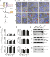
TAT-FADD efficiently delivered inside the cells and retained in the cytoplasm. (A,B) HCT 116 cells were treated with TAT-FADD (TT-FD) at the mentioned concentrations for given time points and, (A) analysis of cell viability and (B) percent LDH release. (CE) HCT 116 cells were transfected with GFP-Caveolin1 for 24 h followed by pre-incubation with MβCD (+MβCD; right panel) for 4 h, further cells were left untreated (0 h) or treated with 5 µM of TAT-FADD for 3–12 h, (C) the internalized TAT-FADD was immunostained with anti-His antibody (His tagged FADD) followed by counterstaining with DAPI and analyzed by confocal microscopy, representative of 25 cells from 3 different fields; scale bar 10 µm and (D) the % localization of TAT-FADD with GFP-Caveolin1 from ‘C’ was calculated with Image J software, (E) analysis of cell viability in the absence (−) and presence (+) of MβCD. (F) HCT 116 cells were treated with 5 µM of TAT-FADD for 3–12 h followed by analysis of cytosolic and nuclear fractions; GAPDH and histone H3 was used as cytosolic and nuclear protein markers, respectively, molecular weight marker left to each blot. The 0 h represents untreated cells. In (A), significance compared between non-treated (NT) and TAT-FADD treated cells; in (B), significance compared between non-treated (0 h) and TAT-FADD treated cells; in (D,E), significance is compared between unprimed (-MβCD; white bars) and primed (+MβCD; black bars) cells treated with TAT-FADD. h, hours; ns, non-significant. Mean  ±  SD; * p  ≤  0.05, ** p  ≤  0.01, and *** p  ≤  0.001.
Figure 3
Figure 3
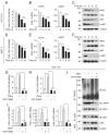
TAT-FADD constituted DISC assembly and induces apoptosis signaling. (A) HCT 116, HeLa, and MCF-7 cells were treated with 5 µM of TAT-FADD for 3–12 h, the His-tagged TAT-FADD was immunoprecipated (IP’ed) using anti-His antibody followed by analysis of DISC assembly proteins procaspase-8 (Pro-C-8) and cFLIPL, molecular weight marker left to each blot. (BF) HCT 116 cells were treated with 5 µM of TAT-FADD for 3–12 h followed by analysis of (B) % apoptotic death by flow cytometry, (C) measurement of caspase-8 (C-8) activity, (D) analysis of apoptosis regulatory proteins, molecular weight marker left to each blot, (E) % change in MMP and (F) measurement of caspase-3 activity. (G,H) HCT 116 cells were transfected with pcDNA3-cFLIPL for 48 h (lane 2 and 5), primed with TNF-α (10 ng/mL) for 12 h (lane 3 and 6) followed by treatment with 5 µM of TAT-FADD for 6 h (lane 4, 5 & 6), (G) analysis of cFLIPL expression by Western blot, molecular weight marker left to each blot and (H) % apoptotic death by a Tali™ image-based cytometer; control represents vector transfected and non-TNF-α-primed cells. In (B,C,E,F), significance compared between non-treated (represents as 0 h) and TAT-FADD-treated cells; in H, significance compared between non-treated (white bars) and TAT-FADD-treated cells (black bars); control in white bar represents a vector transfected and non-TNF-α-primed cells. h, hours; clv, cleaved; MMP, mitochondrial membrane potential. Mean  ±  SD; * p  ≤  0.05, ** p  ≤  0.01 and *** p  ≤  0.001.
Figure 4
Figure 4
TAT-FADD’s pro-apoptotic effect is compared with conventional apoptosis inducers. (A) Schematic diagram representing the target site of proposed apoptosis inducers. (BE) HCT 116 cells were treated with CD 95L (200 ng/mL), TNF-α (50 ng/mL), etoposide (50 µM), HA14-1 (5 µM), protein translational inhibitor cycloheximide (CHX, 5 µg/mL), and TAT-FADD (5 µM) alone for the mentioned time points, (B) The bright field images of cells counterstained with DAPI, post treatments, representative of 150 cells from 3 independent fields, scale bar 5 µm, (C) % apoptotic death by a Tali™ image-based cytometer, (D) % change in MMP and (E) expression of Procaspase-7 and cleavage of PARP by Western blot analysis, molecular weight marker left to each blot. In (C,D), significance compared between non-treated (0 h, white bars) and treated cells (black bars). h, hours; clv, cleaved; CD95-R, CD95 receptor; TNF-R, TNF receptor; MMP, mitochondrial membrane potential. Mean  ±  SD; * p  ≤  0.05, ** p  ≤  0.01, and *** p  ≤  0.001.
Figure 5
Figure 5
TAT-FADD suppresses constitutive and TNF-α-primed NF-κB activation in cancer cells. (AC) HCT 116 cells were treated with 5 µM of TAT-FADD for 3–12 h and (A) NF-κB luciferase activity, (B) mRNA expression of cFLIPL and cIAP2, and (C) expression of NF-κB signaling protein, molecular weight marker left to each blot. (DF) MCF-7 cells were treated with 5 µM of TAT-FADD for 3–12 h and (D) NF-κB luciferase activity, (E) mRNA expression of cFLIPL and cIAP2, and (F) expression of NF-κB signaling protein, molecular weight marker left to each blot. Control represents untreated cells. (GJ) HCT 116 cells were primed with TNF-α (10 ng/mL) for 12 h followed by treatment of 5 µM TAT-FADD for 6 h and (G) NF-κB luciferase activity, (H,I) mRNA expression of (H) Bcl2 and (I) cFLIPL, cIAP2 and RIP1, (J) TRAF2 was immunoprecipitated (IP) followed by binding analysis of ubiquitin, cIAP2, and RIP1 protein by Western blot, molecular weight marker left to each blot. Control represents untreated cells. In (A,B,D,E), significance compared between non-treated (represents as 0 h) and TAT-FADD-treated cells; in (GI), significance is compared between non-treated (white bars) and TNF-α-primed cells (gray bars); and between TNF-α-primed cells and TAT-FADD-treated cells (black and dark gray bars respectively). h, hours; WCL, whole cell lysate. Mean  ±  SD; * p  ≤  0.05, ** p  ≤  0.01, and *** p  ≤  0.001.
Figure 6
Figure 6
TAT-FADD suppresses proinflammatory activation of IL-1β. (A,B) HCT116 cells were treated with 5 µM of TAT-FADD for 3–12 h, (A) mRNA expression of NLRP3, procaspase-1, and pro-IL-1β, and (B) endogenous expression of NLRP3, procaspase-1 (pro Casp-1), and pro IL-1β by Western blot, molecular weight marker left to each blot. The 0 h represents untreated cells. (CE) HCT 116 cells were primed with LPS (100 ng/mL, 12 h) and ATP (5 mM, 2 h) followed by treatment with 5 µM of TAT-FADD for 3–12 h and (C) mRNA expression of pro-IL-1β, (D) expression levels of endogenous NLRP3, procaspase-1, and pro IL-1β by Western blot, molecular weight marker left to each blot, and (E) levels of matured IL-1β was measured by ELISA assay from the culture supernatant. In (C,E) (white bar) and in (D) (lane 1), it represents un-primed and non-treated cells for 12 h taken as controls. In (A), significance is compared between non-treated (represents as 0 h) and TAT-FADD-treated cells; in (C), significance compared between non-treated (white bars) and LPS with ATP-primed cells (gray bars); and between LPS with ATP primed cells treated with TAT-FADD treated cells (black & dark gray bars respectively). h, hours. Mean  ±  SD; ** p  ≤  0.01 and *** p  ≤  0.001.

References

    1. Mouasni S., Tourneur L. Fadd at the crossroads between cancer and inflammation. Trends Immunol. 2018;39:1036–1053. doi: 10.1016/j.it.2018.10.005. - DOI - PubMed
    1. Werner M.H., Wu C., Walsh C.M. Emerging roles for the death adaptor fadd in death receptor avidity and cell cycle regulation. Cell Cycle. 2006;5:2332–2338. doi: 10.4161/cc.5.20.3385. - DOI - PubMed
    1. Scott F.L., Stec B., Pop C., Dobaczewska M.K., Lee J.J., Monosov E., Robinson H., Salvesen G.S., Schwarzenbacher R., Riedl S.J. The fas-fadd death domain complex structure unravels signalling by receptor clustering. Nature. 2009;457:1019–1022. doi: 10.1038/nature07606. - DOI - PMC - PubMed
    1. Lavrik I., Krueger A., Schmitz I., Baumann S., Weyd H., Krammer P.H., Kirchhoff S. The active caspase-8 heterotetramer is formed at the cd95 disc. Cell Death Differ. 2003;10:144–145. doi: 10.1038/sj.cdd.4401156. - DOI - PubMed
    1. Ho P.K., Hawkins C.J. Mammalian initiator apoptotic caspases. FEBS J. 2005;272:5436–5453. doi: 10.1111/j.1742-4658.2005.04966.x. - DOI - PubMed

Substances