. 2020 Sep 18;8(3):20.
doi: 10.3390/jdb8030020.

Reelin Mediates Hippocampal Cajal-Retzius Cell Positioning and Infrapyramidal Blade Morphogenesis

Affiliations

Reelin Mediates Hippocampal Cajal-Retzius Cell Positioning and Infrapyramidal Blade Morphogenesis

Seungshin Ha et al. J Dev Biol. .

Abstract

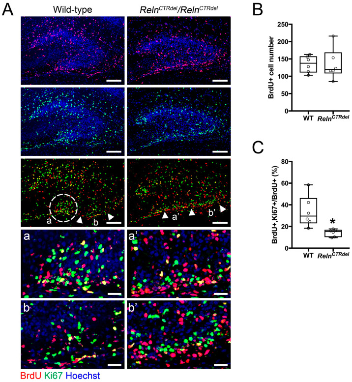
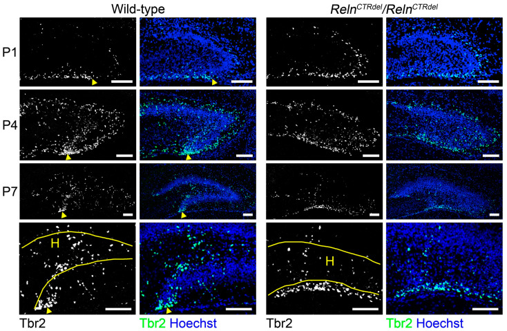
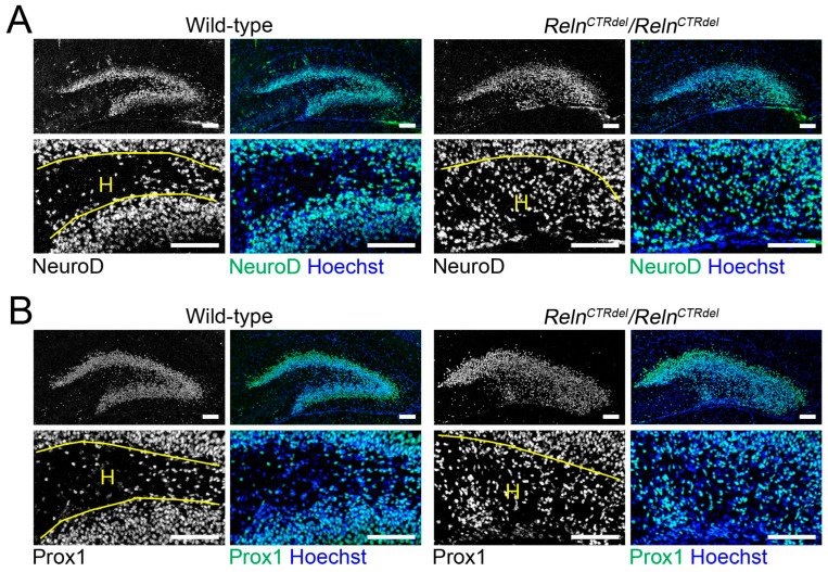
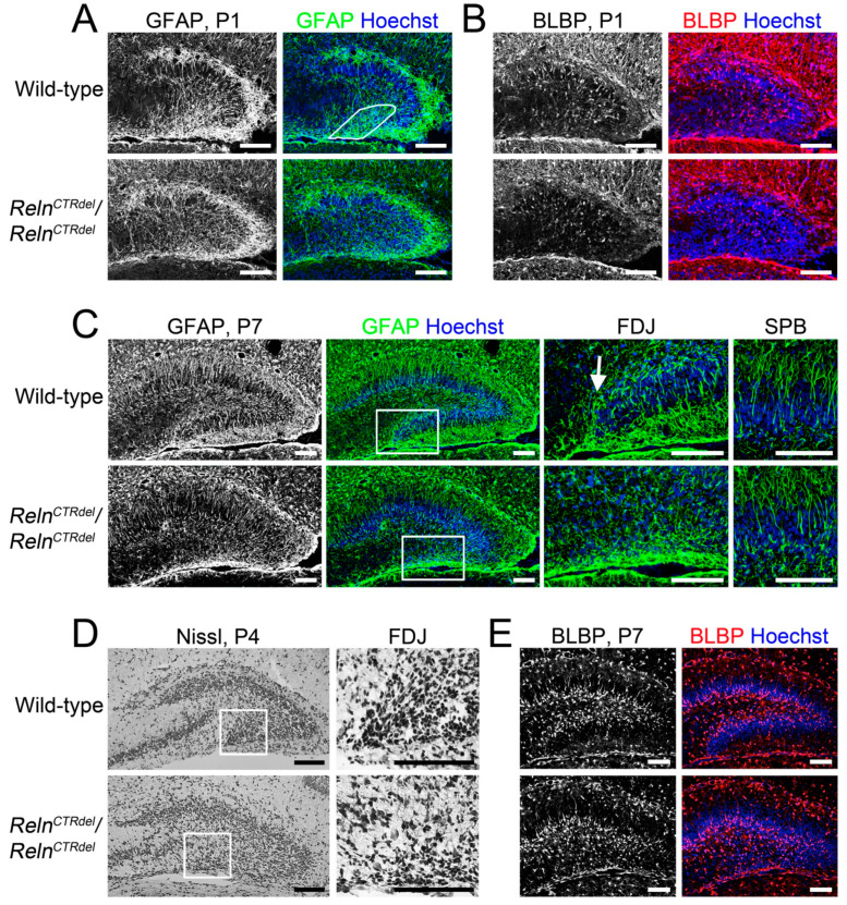
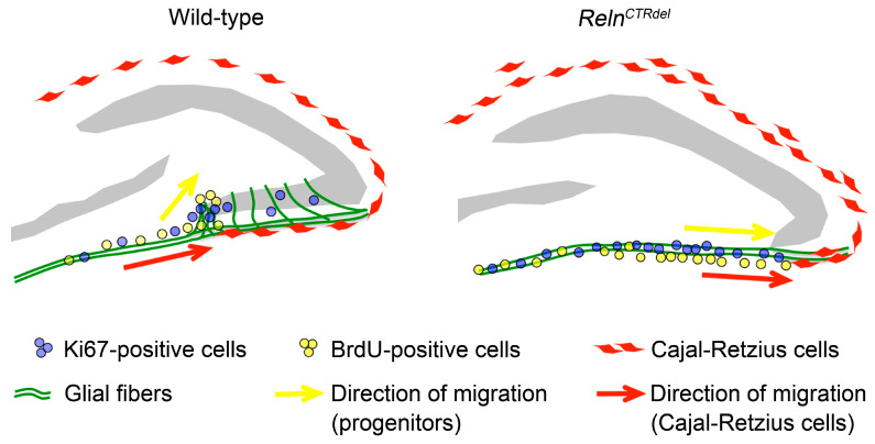
We have previously described hypomorphic reelin (Reln) mutant mice, RelnCTRdel, in which the morphology of the dentate gyrus is distinct from that seen in reeler mice. In the RelnCTRdel mutant, the infrapyramidal blade of the dentate gyrus fails to extend, while the suprapyramidal blade forms with a relatively compact granule neuron layer. Underlying this defect, we now report several developmental anomalies in the RelnCTRdel dentate gyrus. Most strikingly, the distribution of Cajal-Retzius cells was aberrant; Cajal-Retzius neurons were increased in the suprapyramidal blade, but were greatly reduced along the subpial surface of the prospective infrapyramidal blade. We also observed multiple abnormalities of the fimbriodentate junction. Firstly, progenitor cells were distributed abnormally; the "neurogenic cluster" at the fimbriodentate junction was absent, lacking the normal accumulation of Tbr2-positive intermediate progenitors. However, the number of dividing cells in the dentate gyrus was not generally decreased. Secondly, a defect of secondary glial scaffold formation, limited to the infrapyramidal blade, was observed. The densely radiating glial fibers characteristic of the normal fimbriodentate junction were absent in mutants. These fibers might be required for migration of progenitors, which may account for the failure of neurogenic cluster formation. These findings suggest the importance of the secondary scaffold and neurogenic cluster of the fimbriodentate junction in morphogenesis of the mammalian dentate gyrus. Our study provides direct genetic evidence showing that normal RELN function is required for Cajal-Retzius cell positioning in the dentate gyrus, and for formation of the fimbriodentate junction to promote infrapyramidal blade extension.

Keywords: dentate gyrus; hippocampus; migration; neurogenesis; postnatal development; reelin protein.


Conflict of interest statement

The authors declare no conflict of interest.

Figures

Figure A1
Figure A1
Cell death analysis. Activated caspase-3 (green) staining at P4 showed no abnormal increase in cell death in Reln CTRdel hippocampus. Nuclei were stained using Hoechst (blue).
Figure A2
Figure A2
Enlarged images of panels selected from Figure 5 are shown. (A) The right color panel of Figure 5A. Scale bars, 100 µm. (B) The right color panel of Figure 5B. Scale bars, 100 µm. (C) The right panel of Figure 5D. Arrows indicate cells with elongated shape at the FDJ. Scale bars, 50 µm. (D) The right color panel of Figure 5E. Scale bars, 100 µm.
Figure A3
Figure A3
A comparison of the infrapyramidal blade (IPB) malformation between RelnCTRdel and Apoer2null mutants. Apoer2null mutant dentate gyrus also has a truncated IPB although not as severe as RelnCTRdel. This phenotype appears more pronounced in the posterior sections; images of the entire hippocampal region are displayed to show that cross-sections of the similar region were compared. Scale bars, 500 µm.
Figure 1
Figure 1
Lack of the Cajal-Retzius cells in the infrapyramidal blade. (A) The infrapyramidal blade (IPB) of dentate gyrus does not develop in RelnCTRdel, and this coincides with the absence of the RELN-positive cells (gray or red) in the prospective infrapyramidal blade. P7 brains were immunostained with anti-RELN antibodies (n = 2 each). (B) Distribution of p73-expressing cells (gray or green) in the dentate gyrus at P7 (n = 2 each). The number of p73-expressing cells is increased in the suprapyramidal blade (SPB). Nuclei were stained using Hoechst (blue). Scale bars, 250 µm. Arrows indicate the edge of Cajal-Retzius cell-containing regions, which are near the fimbrodentate junction (in the wild type) or the tip of truncated IPB (in the mutants).
Figure 2
Figure 2
Absence of the neurogenic cluster at the fimbriodentate junction. (A) Distribution of the BrdU (red) or Ki67-positive cells (green) in the dentate gyrus at P4, after 24 h from BrdU injection. A cluster of dividing cells is present at the fimbriodentate junction of the wild-type (white circle, a). A RelnCTRdel mutant does not have this cluster (a’). Compared with a corresponding region of the wild type (b), abnormal accumulation of the dividing cells are apparent in the subpial surface of RelnCTRdel (b’). Enlarged images of marked regions (a, a’, b, b’) are shown below. In the mutant, Ki67-positive cells are closely located on top of BrdU-positive cells (a’ and b’). Scale bars, 100 µm. Scale bars in the enlarged images, 25 µm. Nuclei were stained using Hoechst (blue). (B) The number of total BrdU-positive cells is not significantly different (p = 0.8182; boxplot, median ± IQR; whiskers, min and max). (C) The percentage of BrdU/Ki67 double-positive cells was reduced (p = 0.0022; boxplot, median ± IQR; whiskers, min and max). This represents a population of cells that reentered the cell cycle.
Figure 3
Figure 3
Lack of intermediate progenitor accumulation at the fimbriodentate junction. In wild-type mice, Tbr2-positive cells (gray or green) accumulate at the fimbriodentate junction (arrowheads) starting at P1 (n = 2 each) and more clearly at P4 (n = 2 each) and P7 (n = 2 each). This accumulation at FDJ is less apparent in the mutant, and the labeled cells are spread out along the ventricular surface. Enlarged imaged of the hilus at P7 are shown in the lower panel. RelnCTRdel mice have fewer Tbr2-labeled cells in the hilus (H) compared to wild-type mice; it appears as if many cells are still confined to the ventricular surface. Nuclei were stained using Hoechst (blue). Scale bars, 100 µm.
Figure 4
Figure 4
Abnormal distribution of the granule neurons. Granule neuron precursor markers (gray or green), NeuroD (A) and Prox1 (B), staining indicate the presence of abnormal progenitor population in the hilus (H) of RelnCTRdel at P7 (n = 2 each). Nuclei were stained using Hoechst (blue). Enlarged images are shown in lower panels. Scale bars, 100 µm.
Figure 5
Figure 5
Lack of the secondary glial scaffold formation in the IPB. (A) At P1 when the secondary glial scaffold formation begins in the IPB, the mutant mice lack dense GFAP-stained fibers (gray or green, marked region) that are apparent near the fimbriodentate junction of wild-type mice (n = 2 each). Note the premature secondary glial scaffold can be already seen in the SPB of both wild-type and mutant. (B) At P1, BLBP-positive cell bodies (gray or red) appear near the IPB in wild-type mice (n = 2 each). Fewer BLBP-positive cells are found near the IPB of RelnCTRdel mice. (C) Images of GFAP-stained (gray or green) dentate gyrus at P7 (left, n = 2 each). Enlarged images of the boxed fimbriodentate junction (FDJ) regions are shown in the center. In wild-type mice, GFAP-positive fibers are surrounding the growing tip of the infrapyramidal blade at the fimbriodentate junction (arrow). This structure is absent in the mutant. The secondary glia scaffold in the SPB is relatively normal (right, higher-magnification images). (D) At P4, a stream of migrating cells with elongated morphology is absent in the mutant fimbriodentate junction (n = 1 each). Enlarged images of the fimbriodentate junction regions are shown on the right. Nissl stain. (E) At P7, abnormal distribution of BLBP-positive cells (gray or red) in the hilus become more evident (n = 2 each). The cells are directed toward the SPB in the mutant. Nuclei were stained using Hoechst (blue). Scale bars, 100 µm. Enlarged images of (A) (color panel), (B) (color panel), (D) (right panel), and (E) (color panel) can be found in Figure A2.
Figure 6
Figure 6
A model of the infrapyramidal malformation. During the first postnatal week, Cajal-Retzius cells disperse along the subpial surface of the IPB. The progenitors migrate along the dentate migratory stream, then along the densely organized glia scaffold at the fimbriodentate junction, proliferate at the tip and extend the IPB. In RelnCTRdel mutant, Cajal-Retzius cells overmigrate to the SPB and are absent in the subpial surface of the IPB. Without proper RELN signal, the glia scaffolds cannot become organized at the fimbriodentate junction, the progenitor cells fail to migrate toward the hilus, and proliferate within the subpial surface. The mutant dentate gyrus fails to form the outer shell in the IPB, which affects subsequent neurogenesis events in the dentate gyrus.

Similar articles

Cited by

References

    1. Förster E., Zhao S., Frotscher M. Laminating the hippocampus. Nat. Rev. Neurosci. 2006;7:259–268. doi: 10.1038/nrn1882. - DOI - PubMed
    1. Altman J., Bayer S.A. Migration and distribution of two populations of hippocampal granule cell precursors during the perinatal and postnatal periods. J. Comp. Neurol. 1990;301:365–381. doi: 10.1002/cne.903010304. - DOI - PubMed
    1. Li G., Pleasure S.J. Morphogenesis of the Dentate Gyrus: What We Are Learning from Mouse Mutants. Dev. Neurosci. 2005;27:93–99. doi: 10.1159/000085980. - DOI - PubMed
    1. Altman J., Bayer S.A. Mosaic organization of the hippocampal neuroepithelium and the multiple germinal sources of dentate granule cells. J. Comp. Neurol. 1990;301:325–342. doi: 10.1002/cne.903010302. - DOI - PubMed
    1. Hodge R.D., Garcia A.J., Elsen G.E., Nelson B.R., Mussar K.E., Reiner S.L., Ramirez J.-M., Hevner R.F. Tbr2 Expression in Cajal-Retzius Cells and Intermediate Neuronal Progenitors Is Required for Morphogenesis of the Dentate Gyrus. J. Neurosci. 2013;33:4165–4180. doi: 10.1523/JNEUROSCI.4185-12.2013. - DOI - PMC - PubMed