. 2020 Sep 18;21(18):6865.
doi: 10.3390/ijms21186865.

Clearing of Foreign Episomal DNA from Human Cells by CRISPRa-Mediated Activation of Cytidine Deaminases

Affiliations

Clearing of Foreign Episomal DNA from Human Cells by CRISPRa-Mediated Activation of Cytidine Deaminases

Sergey Brezgin et al. Int J Mol Sci. .

Abstract

Restriction of foreign DNA is a fundamental defense mechanism required for maintaining genomic stability and proper function of mammalian cells. APOBEC cytidine deaminases are crucial effector molecules involved in clearing pathogenic DNA of viruses and other microorganisms and improperly localized self-DNA (DNA leakages). Mastering the expression of APOBEC provides the crucial means both for developing novel therapeutic approaches for combating infectious and non-infectious diseases and for numerous research purposes. In this study, we report successful application of a CRISPRa approach to effectively and specifically overexpress APOBEC3A and APOBEC3B deaminases and describe their effects on episomal and integrated foreign DNA. This method increased target gene transcription by >6-50-fold in HEK293T cells. Furthermore, CRISPRa-mediated activation of APOBEC3A/APOBEC3B suppressed episomal but not integrated foreign DNA. Episomal GC-rich DNA was rapidly destabilized and destroyed by CRISPRa-induced APOBEC3A/APOBEC3B, while the remaining DNA templates harbored frequent deaminated nucleotides. To conclude, the CRISPRa approach could be readily utilized for manipulating innate immunity and investigating the effects of the key effector molecules on foreign nucleic acids.

Keywords: APOBECs; CRISPRa; cytidine deaminases; deamination; foreign DNA; innate immunity.


Conflict of interest statement

The authors declare no conflict of interest.

Figures

Figure 1
Figure 1
Design and characteristics of APOBEC3A/APOBEC3B-targeting sgRNAs. The (A) APOBEC3A (A3A1-4) and (B) APOBEC3B (A3B) loci on chromosome 22 shown along with sgRNA target sites, layered with ENCODE regulatory elements and DNase I sensitivity across cell lines indicated as red and black areas, correspondingly. (C) Characteristics (efficacy and off-target effects) of sgRNAs targeting A3A (A3A1-A3A4) and A3B promoters. Efficacy is predicted based on CCTop Broad Institute Online Calculator. MM1–MM4 stand for the number of off-target sites in introns (Int), exons (Ex), and intergenic regions (Intg) (∑ = total number of off-target sites) with the designated number of mismatches (MM1–MM4).
Figure 2
Figure 2
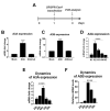
Transactivation of A3A and A3B by CRISPRa. (A) Experimental design. CRISPRa system was transfected into HEK-293T cells the day after seeding. APOBEC mRNA levels were measured 2 days post transfection. (B) A3A and (C) A3B mRNA expression levels upon CRISPRa compared to dCas9p300+NC sgRNA (Mock) or dCas9p300mut+NC sgRNA (mut). (D) Elevation of A3A transcription using CRISPRa with different sgRNAs (A3A1-A3A4). Dynamic analysis of (E) A3A and (F) A3B mRNA levels upon CRISPRa. A3A/A3B mRNA was normalized to GAPDH mRNA. Asterisks indicate statistically significant differences in means. **** p < 0.0001. Mock: dCas9-p300 expressing a plasmid with a non-targeting sgRNA; A3A/A3Bmut: dCas9-p300mut expressing plasmid with the corresponding sgRNA.
Figure 3
Figure 3
CRISPRa transactivation of A3A and A3B decreases GFP signal from an episomal plasmid. (A) Experimental design. HEK-293T cells were co-transfected with a GFP-expressing plasmid and CRISPRa targeting either A3A or A3B and analyzed 5 days post transfection. (B) Bar graph representing percentage of GFP-positive cells in experimental groups. (C) Mean and (D) median fluorescence intensity of transfected cells. (E) Representative FACS plots of untransfected cells (black histogram) and GFP-expressing cells co-transfected with CRISPRa and non-targeting sgRNA (green histogram), A3A-targeting sgRNA (red histogram), or A3B-targeting sgRNA (blue histogram). (F) Semi-quantitative RT-PCR analysis of GFP RNA upon CRISPRa of A3A and A3B genes. GFP mRNA levels were normalized to GAPDH mRNA. (G) Fluorescent images of untransfected, mock-transfected, and CRISPRa-transfected cells. Asterisks indicate statistically significant differences in means. * p < 0.05, ** p < 0.01, **** p < 0.0001.
Figure 4
Figure 4
Analysis of episomal GFP fluorescence in CRISPRa-transfected cells. Histograms of GFP fluorescence in low-signal (R3) to high-signal (R6) populations of (A) untransfected (NT), (B) mock-transfected, (C) A3A-induced, and (D) A3B-induced groups. Semi-quantitative analysis of the % of GFP-positive cells and mean and median GFP fluorescence in (E) mock-treated, (F) A3A-induced, and (G) A3B-induced groups. * p < 0.05, ** p < 0.01, *** p < 0.001.
Figure 5
Figure 5
Decay and deamination of foreign DNA by CRISPRa-mediated activation of A3A and A3B. (A) Decline of GFP-encoding plasmid upon A3A (red bars) and A3B (blue bars) transcriptional activation by CRISPRa. **** p < 0.0001. (B) Deamination of GFP DNA by A3A and A3B measured by 3D-PCR assay.
Figure 6
Figure 6
Toxicity analysis. (A) Cell proliferation and (B) viability were measured in groups transfected with CRISPRa targeting A3A and A3B. CRISPRa with a non-coding sgRNA was used as a mock control. ns: not significant.
Figure 7
Figure 7
CRISPRa-activated A3A and A3B do not affect expression of integrated GFP. (A) Experimental design. HEK-293T cells were transduced with GFP-expressing lentivectors, transfected with CRISPRa systems 5 days later, and analyzed for GFP expression by FACS on day 11. (B) Representative FACS plots of not transduced (black histogram) and GFP-transduced cells co-transfected with CRISPRa and non-targeting sgRNA (green histogram), A3A-targeting sgRNA (red histogram), or A3B-targeting sgRNA (blue histogram). Bar graphs representing (C) percentage of GFP-positive cells, (D) mean and (E) median fluorescence intensity in experimental groups. (F) Fluorescent images of mock-transfected, and CRISPRa-transfected cells. ns: not significant.
Figure 8
Figure 8
Restriction of episomally-encoded foreign DNA by A3A and A3B deaminases overexpressed by CRISPRa. CRISPRa induces specific activation of endogenous A3A and A3B deaminases that effectively suppress and destabilize episomal DNA (resulting in weak GFP signal, reduced GFP plasmid content, and mutated/deaminated GFP DNA). Integrated foreign DNA is less subjected or not subjected (at least in our experimental setting) to the disruptive effects of APOBEC cytidine deaminases. This figure was created in BioRender.

References

    1. Sparrer K.M.J., Gack M.U. Intracellular detection of viral nucleic acids. Curr. Opin. Microbiol. 2015;26:1–9. doi: 10.1016/j.mib.2015.03.001. - DOI - PMC - PubMed
    1. Paludan S.R., Bowie A.G. Immune sensing of DNA. Immunity. 2013;38:870–880. doi: 10.1016/j.immuni.2013.05.004. - DOI - PMC - PubMed
    1. Akira S., Takeda K. Toll-like receptor signalling. Nat. Rev. Immunol. 2004;4:499–511. doi: 10.1038/nri1391. - DOI - PubMed
    1. Sun L., Wu J., Du F., Chen X., Chen Z.J. Cyclic GMP-AMP synthase is a cytosolic DNA sensor that activates the type I interferon pathway. Science. 2013;339:786–791. doi: 10.1126/science.1232458. - DOI - PMC - PubMed
    1. Mussil B., Suspène R., Caval V., Durandy A., Wain-Hobson S., Vartanian J.-P. Genotoxic stress increases cytoplasmic mitochondrial DNA editing by human APOBEC3 mutator enzymes at a single cell level. Sci. Rep. 2019;9:1–11. doi: 10.1038/s41598-019-39245-8. - DOI - PMC - PubMed

LinkOut - more resources