

Beyond Lipid Signaling: Pleiotropic Effects of Diacylglycerol Kinases in Cellular Signaling
- PMID: 32962151
- PMCID: PMC7554708
- DOI: 10.3390/ijms21186861
Beyond Lipid Signaling: Pleiotropic Effects of Diacylglycerol Kinases in Cellular Signaling
Abstract
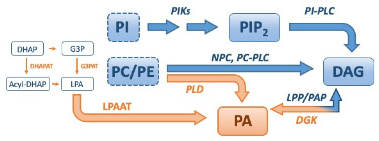
The diacylglycerol kinase family, which can attenuate diacylglycerol signaling and activate phosphatidic acid signaling, regulates various signaling transductions in the mammalian cells. Studies on the regulation of diacylglycerol and phosphatidic acid levels by various enzymes, the identification and characterization of various diacylglycerol and phosphatidic acid-regulated proteins, and the overlap of different diacylglycerol and phosphatidic acid metabolic and signaling processes have revealed the complex and non-redundant roles of diacylglycerol kinases in regulating multiple biochemical and biological networks. In this review article, we summarized recent progress in the complex and non-redundant roles of diacylglycerol kinases, which is expected to aid in restoring dysregulated biochemical and biological networks in various pathological conditions at the bed side.
Keywords: diacylglycerol; diacylglycerol kinase; lipid signaling; phosphatidic acid; tissue microenvironment; tumor microenvironment.
Conflict of interest statement
The authors declare no conflict of interest. The funders had no role in the design of the study; in the collection, analyses, or interpretation of data; in the writing of the manuscript, or in the decision to publish the results.
Figures


Similar articles
-
Signaling roles of diacylglycerol kinases.J Cell Biochem. 2006 Feb 15;97(3):474-84. doi: 10.1002/jcb.20704. J Cell Biochem. 2006. PMID: 16288460 Review.
-
Diacylglycerol kinases: regulation and signaling roles.Thromb Haemost. 2002 Dec;88(6):912-8. Thromb Haemost. 2002. PMID: 12529738 Review.
-
Diacylglycerol, phosphatidic acid, and the converting enzyme, diacylglycerol kinase, in the nucleus.Biochim Biophys Acta. 2006 May-Jun;1761(5-6):535-41. doi: 10.1016/j.bbalip.2006.04.001. Epub 2006 Apr 20. Biochim Biophys Acta. 2006. PMID: 16731035 Review.
-
Diacylglycerol kinase ζ: at the crossroads of lipid signaling and protein complex organization.Prog Lipid Res. 2012 Jan;51(1):1-10. doi: 10.1016/j.plipres.2011.10.001. Epub 2011 Nov 2. Prog Lipid Res. 2012. PMID: 22067957 Review.
-
Regulation of DGK-theta.J Cell Physiol. 2009 Sep;220(3):548-52. doi: 10.1002/jcp.21813. J Cell Physiol. 2009. PMID: 19472209 Review.
Cited by
-
Diacylglycerol Kinases and Its Role in Lipid Metabolism and Related Diseases.Int J Mol Sci. 2024 Dec 9;25(23):13207. doi: 10.3390/ijms252313207. Int J Mol Sci. 2024. PMID: 39684917 Free PMC article. Review.
-
Diacylglycerol kinase epsilon protects against renal ischemia/reperfusion injury in mice through Krüppel-like factor 15/klotho pathway.Ren Fail. 2022 Dec;44(1):902-913. doi: 10.1080/0886022X.2022.2079524. Ren Fail. 2022. PMID: 35616094 Free PMC article.
-
Store-Operated Ca2+ Entry Is Up-Regulated in Tumour-Infiltrating Lymphocytes from Metastatic Colorectal Cancer Patients.Cancers (Basel). 2022 Jul 7;14(14):3312. doi: 10.3390/cancers14143312. Cancers (Basel). 2022. PMID: 35884372 Free PMC article.
-
Integrated Network Pharmacology and Lipidomics to Reveal the Inhibitory Effect of Qingfei Oral Liquid on Excessive Autophagy in RSV-Induced Lung Inflammation.Front Pharmacol. 2021 Dec 1;12:777689. doi: 10.3389/fphar.2021.777689. eCollection 2021. Front Pharmacol. 2021. PMID: 34925035 Free PMC article.
-
Diacylglycerol Kinases in Signal Transduction.Int J Mol Sci. 2022 Jul 29;23(15):8423. doi: 10.3390/ijms23158423. Int J Mol Sci. 2022. PMID: 35955558 Free PMC article.
References
-
- Kanoh H., Kondoh H., Ono T. Diacylglycerol kinase from pig brain. Purification and phospholipid dependencies. J. Biol. Chem. 1983;258:1767–1774. - PubMed
Publication types
MeSH terms
Substances
Grants and funding
LinkOut - more resources
Full Text Sources
Other Literature Sources
