

Human Pluripotent Stem Cell-Derived Neural Cells as a Relevant Platform for Drug Screening in Alzheimer's Disease
- PMID: 32962164
- PMCID: PMC7558359
- DOI: 10.3390/ijms21186867
Human Pluripotent Stem Cell-Derived Neural Cells as a Relevant Platform for Drug Screening in Alzheimer's Disease
Abstract
Extracellular amyloid-beta deposition and intraneuronal Tau-laden neurofibrillary tangles are prime features of Alzheimer's disease (AD). The pathology of AD is very complex and still not fully understood, since different neural cell types are involved in the disease. Although neuronal function is clearly deteriorated in AD patients, recently, an increasing number of evidences have pointed towards glial cell dysfunction as one of the main causative phenomena implicated in AD pathogenesis. The complex disease pathology together with the lack of reliable disease models have precluded the development of effective therapies able to counteract disease progression. The discovery and implementation of human pluripotent stem cell technology represents an important opportunity in this field, as this system allows the generation of patient-derived cells to be used for disease modeling and therapeutic target identification and as a platform to be employed in drug discovery programs. In this review, we discuss the current studies using human pluripotent stem cells focused on AD, providing convincing evidences that this system is an excellent opportunity to advance in the comprehension of AD pathology, which will be translated to the development of the still missing effective therapies.
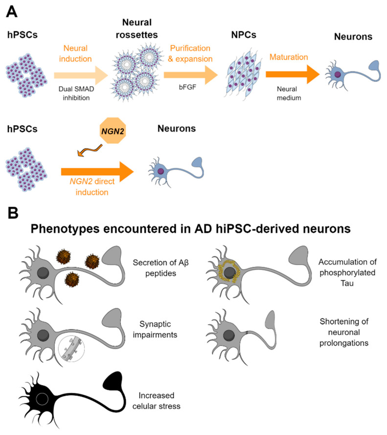
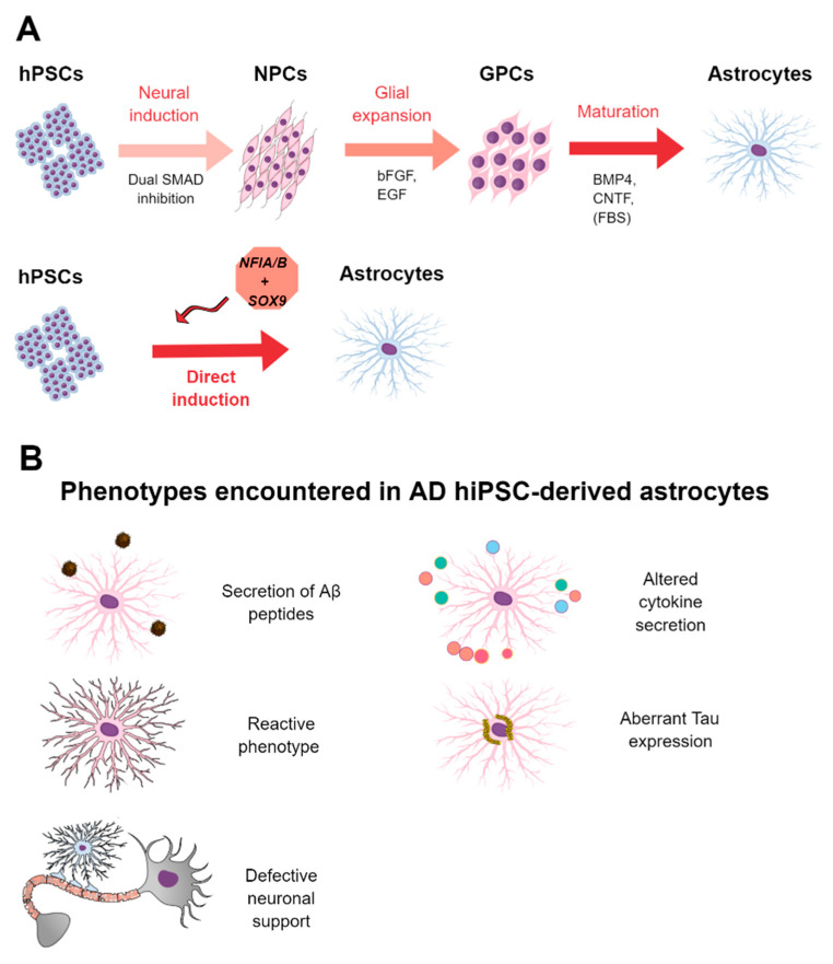
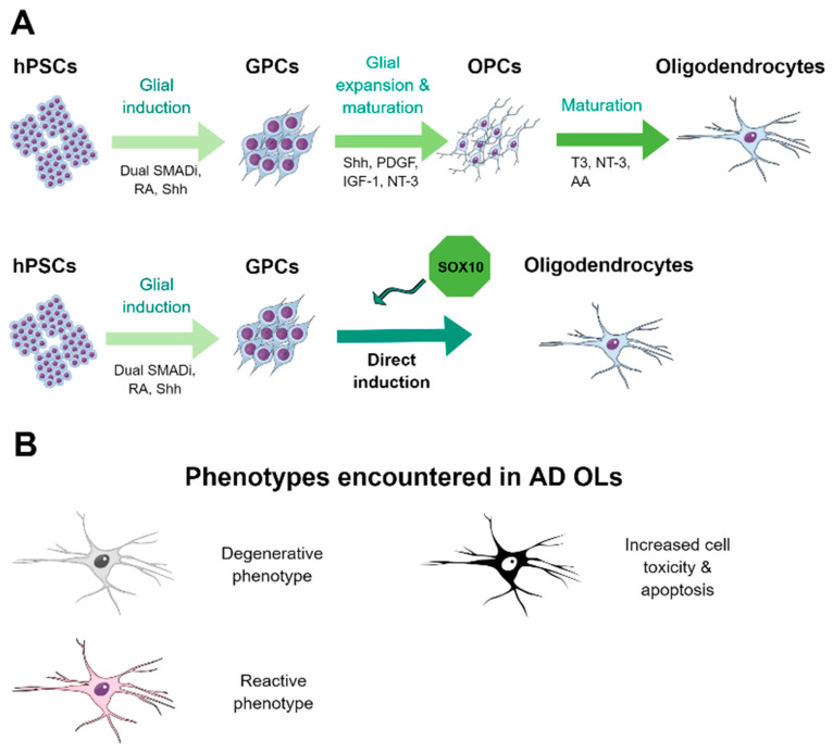
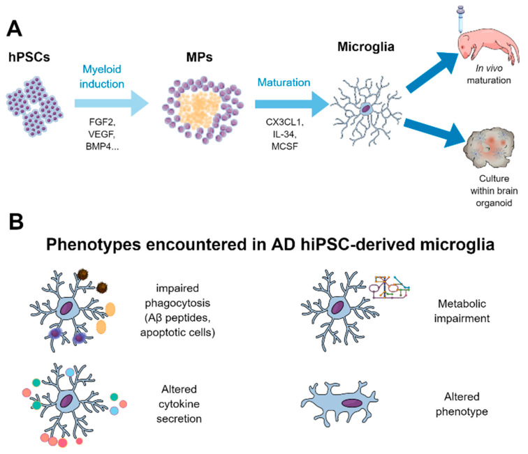
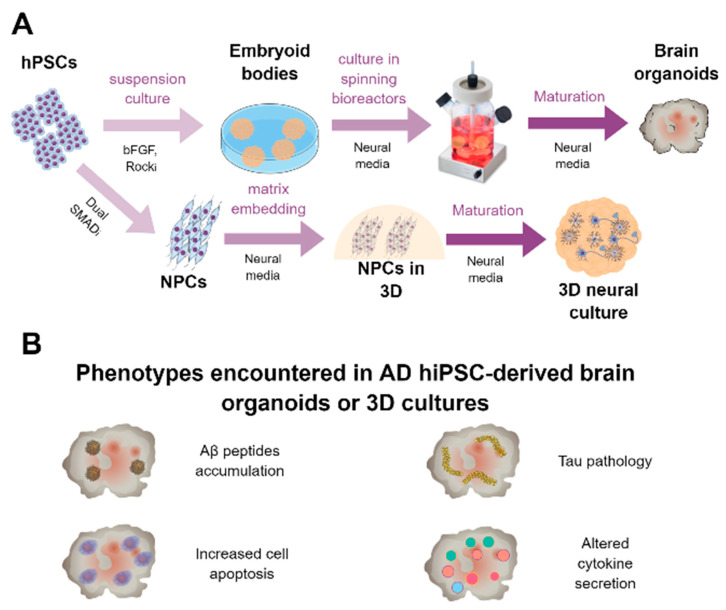
Keywords: 3D cultures; Alzheimer’s disease; astrocytes; brain organoids; disease modeling; human induced pluripotent stem cells (hiPSCs); microglia; oligodendrocytes.
Conflict of interest statement
The authors declare no conflict of interest.
Figures
Similar articles
-
Modeling amyloid beta and tau pathology in human cerebral organoids.Mol Psychiatry. 2018 Dec;23(12):2363-2374. doi: 10.1038/s41380-018-0229-8. Epub 2018 Aug 31. Mol Psychiatry. 2018. PMID: 30171212 Free PMC article.
-
A three-dimensional human neural cell culture model of Alzheimer's disease.Nature. 2014 Nov 13;515(7526):274-8. doi: 10.1038/nature13800. Epub 2014 Oct 12. Nature. 2014. PMID: 25307057 Free PMC article.
-
Extracellular vesicles from human-induced pluripotent stem cell-derived neural stem cells alleviate proinflammatory cascades within disease-associated microglia in Alzheimer's disease.J Extracell Vesicles. 2024 Nov;13(11):e12519. doi: 10.1002/jev2.12519. J Extracell Vesicles. 2024. PMID: 39499013 Free PMC article.
-
Human iPSC-Derived Neural Models for Studying Alzheimer's Disease: from Neural Stem Cells to Cerebral Organoids.Stem Cell Rev Rep. 2022 Feb;18(2):792-820. doi: 10.1007/s12015-021-10254-3. Epub 2022 Feb 2. Stem Cell Rev Rep. 2022. PMID: 35107767 Free PMC article. Review.
-
Alzheimer's disease and synapse Loss: What can we learn from induced pluripotent stem Cells?J Adv Res. 2023 Dec;54:105-118. doi: 10.1016/j.jare.2023.01.006. Epub 2023 Jan 14. J Adv Res. 2023. PMID: 36646419 Free PMC article. Review.
Cited by
-
Exploring the promising potential of induced pluripotent stem cells in cancer research and therapy.Mol Cancer. 2023 Nov 28;22(1):189. doi: 10.1186/s12943-023-01873-0. Mol Cancer. 2023. PMID: 38017433 Free PMC article. Review.
-
Comparison of Extracellular Vesicles from Induced Pluripotent Stem Cell-Derived Brain Cells.Int J Mol Sci. 2024 Mar 22;25(7):3575. doi: 10.3390/ijms25073575. Int J Mol Sci. 2024. PMID: 38612385 Free PMC article.
-
Comprehensive characterization of the neurogenic and neuroprotective action of a novel TrkB agonist using mouse and human stem cell models of Alzheimer's disease.Stem Cell Res Ther. 2024 Jul 6;15(1):200. doi: 10.1186/s13287-024-03818-w. Stem Cell Res Ther. 2024. PMID: 38971770 Free PMC article.
-
The effects and potential of microglial polarization and crosstalk with other cells of the central nervous system in the treatment of Alzheimer's disease.Neural Regen Res. 2023 May;18(5):947-954. doi: 10.4103/1673-5374.355747. Neural Regen Res. 2023. PMID: 36254973 Free PMC article. Review.
-
Advances in Recapitulating Alzheimer's Disease Phenotypes Using Human Induced Pluripotent Stem Cell-Based In Vitro Models.Brain Sci. 2022 Apr 26;12(5):552. doi: 10.3390/brainsci12050552. Brain Sci. 2022. PMID: 35624938 Free PMC article. Review.
References
-
- Winblad B., Amouyel P., Andrieu S., Ballard C., Brayne C., Brodaty H., Cedazo-Minguez A., Dubois B., Edvardsson D., Feldman H., et al. Defeating Alzheimer’s disease and other dementias: A priority for European science and society. Lancet Neurol. 2016;15:455–532. doi: 10.1016/S1474-4422(16)00062-4. - DOI - PubMed
Publication types
MeSH terms
Substances
Grants and funding
- PI18/01557; PI18/01556;/Instituto de Salud Carlos III
- CB06/05/1116; CB06/05/0094/Centro de Investigación Biomédica en Red sobre Enfermedades Neurodegenerativas
- UMA18-FEDERJA-211; PY18-RT-2233/Consejería de Economía, Innovación, Ciencia y Empleo, Junta de Andalucía
- PI-0276-2018/Consejeria de Salud, Junta de Andalucia
- PPIT.UMA.B1.2017/26;/Universidad de Málaga
