

Taurine Inhibits Glucocorticoid-Induced Bone Mitochondrial Injury, Preventing Osteonecrosis in Rabbits and Cultured Osteocytes
- PMID: 32962196
- PMCID: PMC7555938
- DOI: 10.3390/ijms21186892
Taurine Inhibits Glucocorticoid-Induced Bone Mitochondrial Injury, Preventing Osteonecrosis in Rabbits and Cultured Osteocytes
Abstract
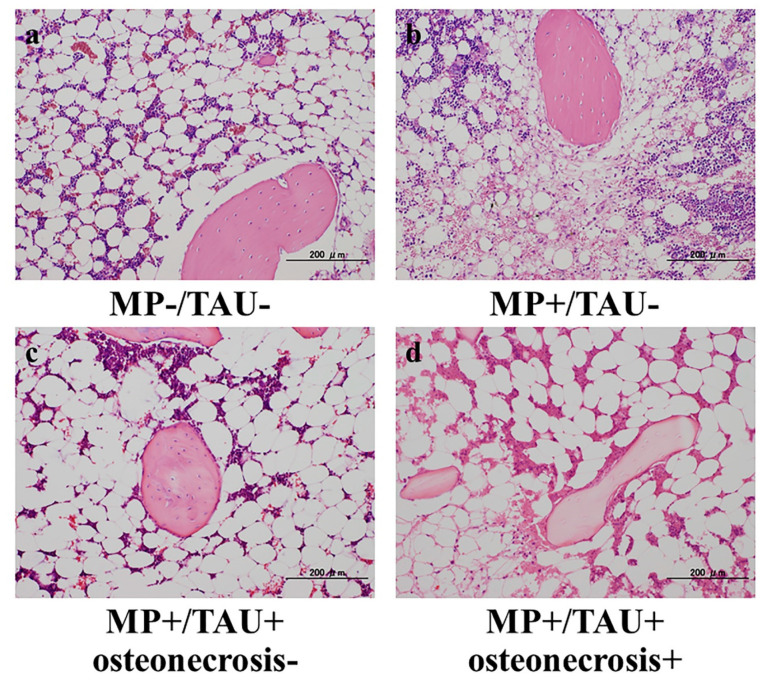
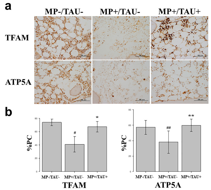
Mitochondrial injury has recently been implicated in the pathogenesis of glucocorticoid-induced osteonecrosis. Using cultured osteocytes and a rabbit model, we investigated the possibility that taurine (TAU), which is known to play a role in the preservation of mitochondrial function, might also prevent the development of osteonecrosis. To reduplicate the intraosseous environment seen in glucocorticoid-induced osteonecrosis, dexamethasone (Dex) was added to MLO-Y4 cultured in 1% hypoxia (H-D stress environment). An in vitro study was conducted in which changes in mitochondrial transcription factor A (TFAM), a marker of mitochondrial function, and ATP5A produced by mitochondria, induced by the presence/absence of taurine addition were measured. To confirm the effect of taurine in vivo, 15 Japanese White rabbits were administered methylprednisolone (MP) 20 mg/kg as a single injection into the gluteus muscle (MP+/TAU- group), while for 5 consecutive days from the day of MP administration, taurine 100 mg/kg was administered to 15 animals (MP+/TAU+ group). As a control 15 untreated rabbits were also studied. The rabbits in each of the groups were sacrificed on the 14th day after glucocorticoid administration, and the bilateral femora were harvested. Histopathologically, the incidence of osteonecrosis was quantified immunohistochemically by quantifying TFAM and ATP5A expression. In the rabbits exposed to an H-D stress environment and in MP+/TAU- group, TFAM and ATP5A expression markedly decreased. With addition of taurine in the in vitro and in vivo studies, the expression of TFAM and ATP5A was somewhat decreased as compared with Dex-/hypoxia- or MP-/TAU- group, while improvement was noted as compared with Dex+/hypoxia+ or MP+/TAU- group. In rabbits, the incidence of osteonecrosis was 80% in MP+/TAU- group, in contrast to 20% in the taurine administered group (MP+/TAU+), representing a significant decrease. Since taurine was documented to exert a protective effect on mitochondrial function by inhibiting the mitochondrial dysfunction associated with glucocorticoid administration, we speculated that it might also indirectly help to prevent the development of osteonecrosis in this context. Since taurine is already being used clinically, we considered that its clinical application would also likely be smooth.
Keywords: glucocorticoid; mitochondrial function; osteonecrosis; taurine.
Conflict of interest statement
The authors declare no conflict of interest.
Figures
Similar articles
-
Mitochondrial Transcription Factor A added to Osteocytes in a Stressed Environment has a Cytoprotective Effect.Int J Med Sci. 2020 May 23;17(9):1293-1299. doi: 10.7150/ijms.45335. eCollection 2020. Int J Med Sci. 2020. PMID: 32547324 Free PMC article.
-
Resistance of bone marrow mesenchymal stem cells in a stressed environment - Comparison with osteocyte cells.Int J Med Sci. 2021 Jan 21;18(6):1375-1381. doi: 10.7150/ijms.52104. eCollection 2021. Int J Med Sci. 2021. PMID: 33628093 Free PMC article.
-
Prevention of glucocorticoid-associated osteonecrosis by intravenous administration of mesenchymal stem cells in a rabbit model.BMC Musculoskelet Disord. 2017 Nov 21;18(1):480. doi: 10.1186/s12891-017-1837-1. BMC Musculoskelet Disord. 2017. PMID: 29162088 Free PMC article.
-
Glucocorticoid-induced osteonecrosis.Endocrine. 2012 Apr;41(2):183-90. doi: 10.1007/s12020-011-9580-0. Epub 2011 Dec 15. Endocrine. 2012. PMID: 22169965 Free PMC article. Review.
-
Role of apoptosis in glucocorticoid-induced osteoporosis and osteonecrosis.Crit Rev Eukaryot Gene Expr. 2003;13(2-4):221-35. Crit Rev Eukaryot Gene Expr. 2003. PMID: 14696969 Review.
Cited by
-
Taurine as a Natural Antioxidant: From Direct Antioxidant Effects to Protective Action in Various Toxicological Models.Antioxidants (Basel). 2021 Nov 24;10(12):1876. doi: 10.3390/antiox10121876. Antioxidants (Basel). 2021. PMID: 34942978 Free PMC article. Review.
-
Identification and analysis of mitochondria-related central genes in steroid-induced osteonecrosis of the femoral head, along with drug prediction.Front Endocrinol (Lausanne). 2024 Feb 7;15:1341366. doi: 10.3389/fendo.2024.1341366. eCollection 2024. Front Endocrinol (Lausanne). 2024. PMID: 38384969 Free PMC article.
-
The role of taurine in bone metabolism.iScience. 2025 Aug 5;28(9):113257. doi: 10.1016/j.isci.2025.113257. eCollection 2025 Sep 19. iScience. 2025. PMID: 40894901 Free PMC article. Review.
-
Mitochondrial maintenance as a novel target for treating steroid-induced osteonecrosis of femoral head: a narrative review.EFORT Open Rev. 2024 Nov 8;9(11):1013-1022. doi: 10.1530/EOR-24-0023. EFORT Open Rev. 2024. PMID: 39513701 Free PMC article. Review.
-
Research of Pathogenesis and Novel Therapeutics in Arthritis 2.0.Int J Mol Sci. 2020 Oct 30;21(21):8125. doi: 10.3390/ijms21218125. Int J Mol Sci. 2020. PMID: 33143215 Free PMC article.
References
MeSH terms
Substances
LinkOut - more resources
Full Text Sources
Medical
Research Materials
Miscellaneous
