

The Emergence of a vv + MDV Can Break through the Protections Provided by the Current Vaccines
- PMID: 32962247
- PMCID: PMC7551601
- DOI: 10.3390/v12091048
The Emergence of a vv + MDV Can Break through the Protections Provided by the Current Vaccines
Abstract
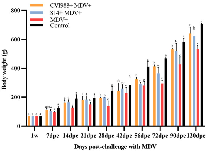
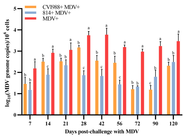
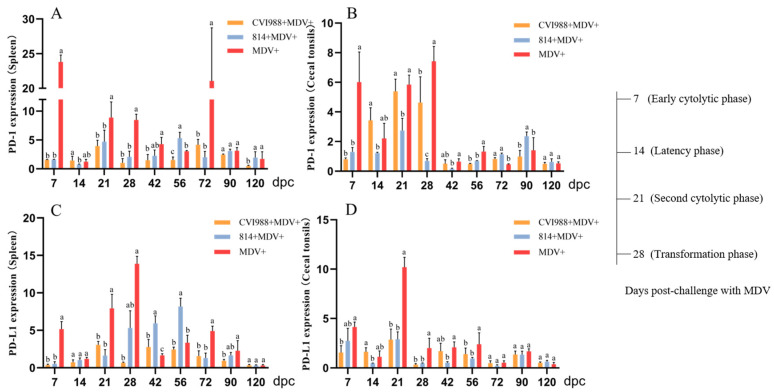
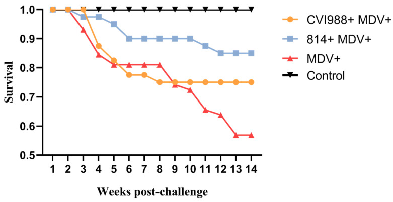
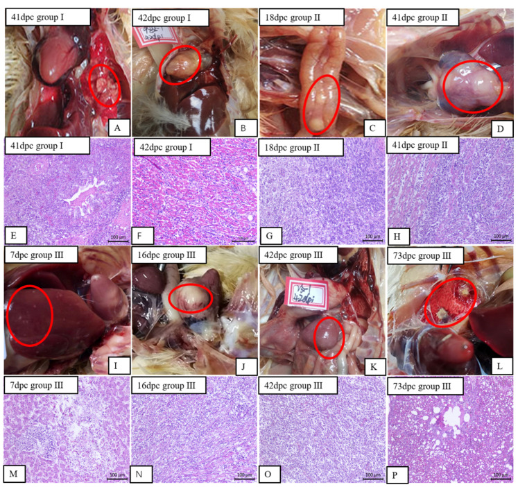
Marek's disease (MD) is an infectious malignant T-cell lymphoma proliferative disease caused by Marek's disease virus (MDV). In recent years, the emergence of very virulent (vv) and/or very virulent plus (vv +) strains of MDV in the field has been suggested as one of the causes of vaccination failure. The pathogenicity of the MDV strain GX18NNM4, isolated from a clinical outbreak in a broiler breeder flock that was vaccinated with CVI988/Rispens, was investigated. In the vaccination-challenge test, GX18NNM4 was able to break through the protections provided by the vaccines CVI988 and 814. It also significantly reduced body weight gain and caused marked gross lesions and a large area of infiltration of neoplastic lymphocyte cells in the heart, liver, pancreas, etc. of the infected birds. In addition, the expressions of programmed death 1 (PD-1) and its ligand, programmed death ligand 1 (PD-L1), in the spleens and cecal tonsils (CTs) of the unvaccinated challenged birds were significantly increased compared to those in the vaccinated challenged birds, indicating that the PD-1/PD-L1 pathway is related to immune evasion mechanisms. The results showed that the GX18NNM4 strain could cause severe immunosuppression and significantly decrease the protections provided by the current commercial vaccines, thus showing GX18NNM4 to be a vv + MDV strain.
Keywords: Marek’s disease virus; immunosuppression; pathogenicity analysis; protection index; vv + MDV.
Conflict of interest statement
The authors declare no conflict of interest.
Figures

References
-
- Nolan L.K., Swayne D.E., Saif Y.M., Fadly A.M., Glisson J.R., Mcdougald L.R. Diseases of Poultry. Wiley; Hoboken, NJ, USA: 2016. pp. 513–673.
-
- Morrow C., Fehler F. 5-Marek’s disease: A worldwide problem. In: Davison F., Nair V., editors. Marek’s Disease. Academic Press; Oxford, UK: 2004. pp. 49–61.
Publication types
MeSH terms
Substances
LinkOut - more resources
Full Text Sources
Research Materials
