

Gastric schwannoma: a case report and literature review
- PMID: 32962485
- PMCID: PMC7518005
- DOI: 10.1177/0300060520957828
Gastric schwannoma: a case report and literature review
Abstract
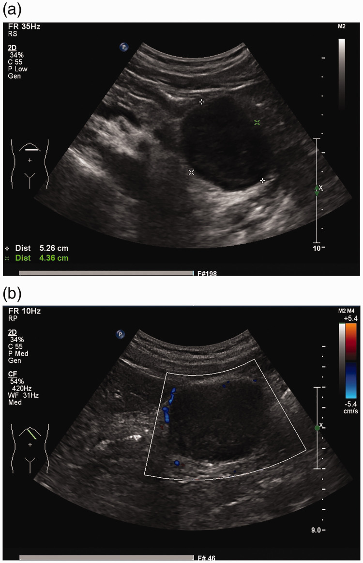
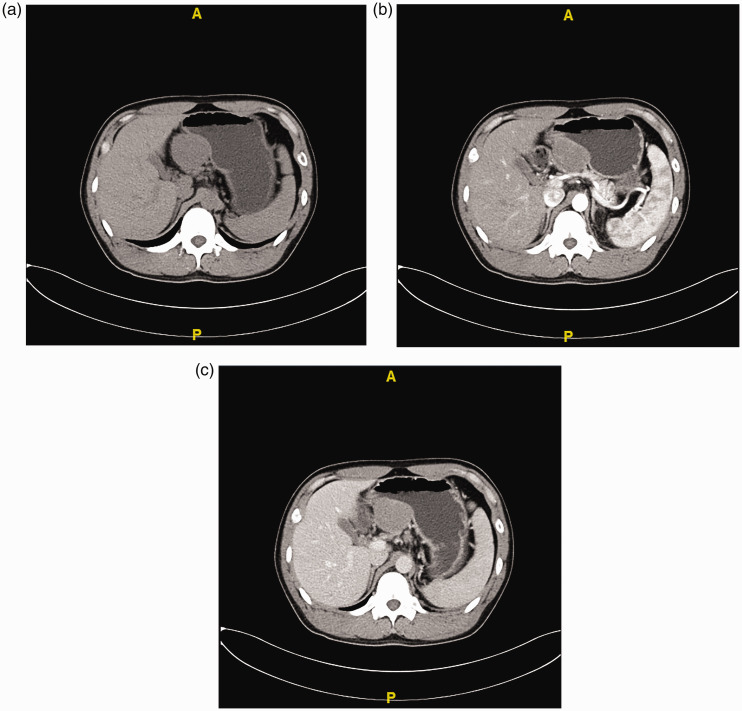
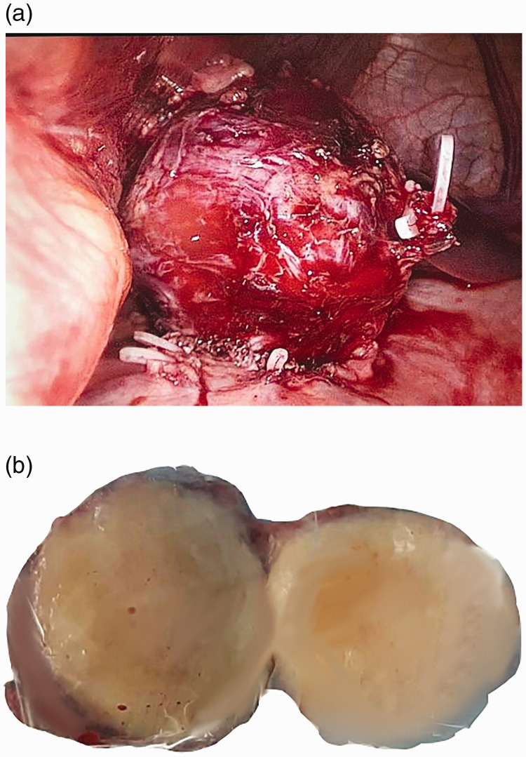
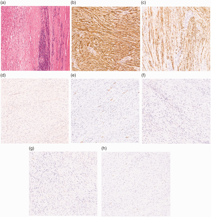
Background: Gastric schwannoma is a rarely seen gastric tumor accounting for only 0.2% of all gastric tumors. It is difficult to distinguish a gastric schwannoma from other gastric tumors preoperatively.Case presentation: A 30-year-old man with no significant medical history or physical examination findings presented with a 1-month history of right upper abdominal discomfort. The preoperative diagnosis was a gastrointestinal stromal tumor, but the postoperative pathologic and immunohistochemical examinations confirmed a gastric schwannoma. The patient underwent laparoscopic wedge resection of the stomach without additional postoperative treatment, and his postoperative recovery was uneventful. No recurrence or metastasis was found at the 2-year follow-up examination.
Conclusion: Although gastric schwannomas are usually not malignant, they are difficult to distinguish from other malignant stromal tumors preoperatively. Surgical resection should be recommended when a schwannoma is malignant or considered to be at risk of becoming malignant.
Keywords: Schwannoma; gastric schwannoma; gastroenterology and hepatology; gastrointestinal stromal tumor; general surgery; laparoscopic wedge resection.
Figures
References
-
- Daimaru Y, Kido H, Hashimoto H, et al. Benign schwannoma of the gastrointestinal tract: a clinicopathologic and immunohistochemical study. Hum Pathol 1988; 19: 257–264. - PubMed
-
- Prevot S, Bienvenu L, Vaillant JC, et al. Benign schwannoma of the digestive tract: a clinicopathologic and immunohistochemical study of five cases, including a case of esophageal tumor. Am J Surg Pathol 1999; 23: 431–436. - PubMed
Publication types
MeSH terms
LinkOut - more resources
Full Text Sources
Medical
