

Saikosaponin D inhibits proliferation and induces apoptosis of non-small cell lung cancer cells by inhibiting the STAT3 pathway
- PMID: 32962498
- PMCID: PMC7780581
- DOI: 10.1177/0300060520937163
Saikosaponin D inhibits proliferation and induces apoptosis of non-small cell lung cancer cells by inhibiting the STAT3 pathway
Abstract
Objective: To study the effects of saikosaponin D (SSD) on proliferation and apoptosis in human non-small cell lung cancer cell lines, and to explore underlying mechanisms.
Methods: Following treatment with saikosaponin D, A549 and H1299 cells were assessed for anti-proliferation effects using cell cycle kit-8 assays, changes in nuclear morphology using 4',6-diamidino-2-phenylindole (DAPI) staining, and cell apoptosis using annexin V/propidium iodide double staining. Proliferation- and apoptosis-related proteins were detected by immunoblotting.
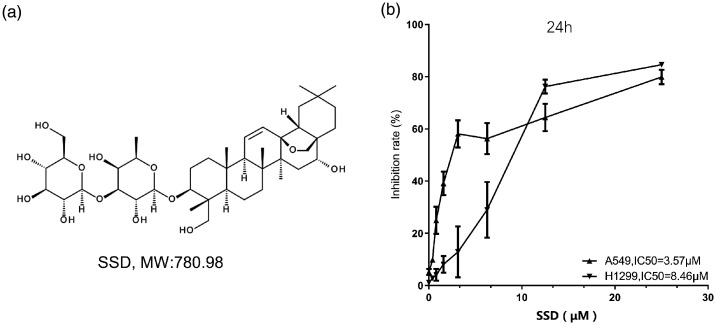
Results: Saikosaponin D had dose-dependent inhibitory effects on A549 cells (IC50, 3.57 µM) and H1299 cells (IC50, 8.46 µM). DAPI staining revealed decreased cell numbers, and most H1299 cells became round after treatment with 20 µM saikosaponin D. As saikosaponin D concentration increased, the proportions of cells in G0/G1 phase, and cells undergoing apoptosis, increased. Levels of phosphorylated p44/42 and signal transducer and activator of transcription (STAT)3 were significantly downregulated in both cell lines, while total STAT3 levels were not significantly affected. The cleaved form of caspase 3 was significantly upregulated.
Conclusions: Saikosaponin D inhibits proliferation, inducing cell cycle arrest and apoptosis, in lung cancer cells in a dose-dependent manner, possibly through inhibition of STAT3 phosphorylation and activation of caspase 3.
Keywords: STAT3 pathway; Saikosaponin D; apoptosis; lung cancer.
Figures


References
-
- Remon J, Ahn MJ, Girard N, et al. Advanced-stage non-small cell lung cancer: advances in thoracic oncology 2018. J Thorac Oncol 2019; 14: 1134–1155. - PubMed
-
- Saranya K, Sreejith K. and Ajaykumar . Comparison of quality of life of patients on treatment with cisplatin and gemcitabine, carboplatin and gemcitabine, carboplatin and paclitaxel, carboplatin and pemetrexed for non-small cell lung cancer. J Oncol Pharm Pract 2019; 25: 1853–1859. - PubMed
-
- Kujtan L, Subramanian J. Epidermal growth factor receptor tyrosine kinase inhibitors for the treatment of non-small cell lung cancer. Expert Rev Anticancer Ther 2019; 19: 547–559. - PubMed
-
- Xie JY, Di HY, Li H, et al. Bupleurum chinense DC polysaccharides attenuates lipopolysaccharide-induced acute lung injury in mice. Phytomedicine 2012; 19: 130–137. - PubMed
MeSH terms
Substances
LinkOut - more resources
Full Text Sources
Medical
Research Materials
Miscellaneous
