Case Reports
. 2020 Sep 22;20(1):251.
doi: 10.1186/s12890-020-01287-4.

Multiple endo bronchial lipoma: a rare case report

Affiliations
Case Reports

Multiple endo bronchial lipoma: a rare case report

Shunjin Zhao et al. BMC Pulm Med. .

Abstract

Background: Endobronchial lipoma is an extremely rare benign tumor, which is generally located in the first three subdivisions of the tracheobronchial tree. According to the existing literature, all endobronchial lipomas are single (one per patient). Here, we report a rare case in which the patient presented with two endobronchial lipomas in the same patient, and underwent a bronchoscopic tumor resection in the left main bronchus and the left lower bronchus. Both tumors were pathologically confirmed as endobronchial lipoma.

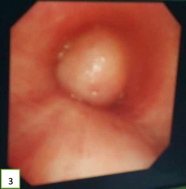
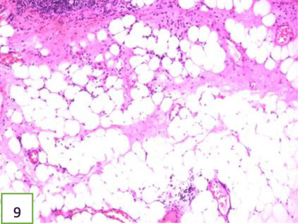
Case presentation: A 52-year-old Chinese man presented at the clinic reporting a mild cough with yellow color sputum and exertional dyspnea for 2 weeks. He was a heavy smoker (45 pack-years). Chest auscultation demonstrated faint wheezing in left lower lobe. Computed tomography (CT) revealed two low-density endobronchial masses located in the middle segment of the left main bronchus and the posterior basilar segmental bronchus of the left lower lobe. The neoplasms measured a CT-attenuation value of -70HU, -98HU in density with air trapping and atelectasis in the segmental bronchus of the left lower lobe. The patient underwent interventional bronchoscopic management to remove the neoplasms by using an electrosurgical snare, cryotherapy, and electrocautery. The locations of the neoplasms were confirmed at the left main bronchus and the superior segment of the left lower lobe during bronchoscopic intervention. Histopathological examination confirmed that both tissues were consistent with lipomas. After 18 months of follow-up, the patient was free of symptoms and CT revealed that bronchiectasia remained in the superior segment of the left lower lobe; however, no mass lesion was present in the left bronchus.

Conclusions: This case suggests that an endobronchial lipoma can present as multiple lesions, and both proximal and distal types can simultaneously occur in the same patient. Thus, these findings help us further understand the biology of endobronchial lipomas.

Keywords: Bronchoscopy; Case report; Endobronchial lipoma; Lipoma.


Conflict of interest statement

The authors declare that they have no competing interests.

Figures

Fig. 1
Fig. 1
CT showing a low-density mass obstructing the left main bronchus, the CT value is -70HU
Fig. 2
Fig. 2
CT suggesting low-density mass obstructing the left lower lobe, the CT value is -98HU
Fig. 3
Fig. 3
A spherical neoplasm growing in the left main bronchus with a smooth, soft, and red appearance
Fig. 4
Fig. 4
Neoplasm located in left main bronchus, about 2.0 × 1.3 × 0.9 cm in size, with an oval polypoid shape
Fig. 5
Fig. 5
The left lower lobe bronchus is blocked by a neoplasm. Its surface is smooth, soft, has a light-yellow appearance, and is dumbbell shaped
Fig. 6
Fig. 6
Image of left main bronchial tumor after resection
Fig. 7
Fig. 7
Five pieces of neoplasia tissues that have been removed from the left main bronchus and the lower left bronchus
Fig. 8
Fig. 8
Image of the left lower lobe bronchial after tumor resection
Fig. 9
Fig. 9
Histology of the neoplasm at low magnification demonstrating the pseudo-stratified ciliated columnar epithelium overlying the mature adipocytes and little fibrous tissue. The neoplasm in the left main bronchus is consistent with lipoma. Hematoxylin and eosin (HE) staining
Fig. 10
Fig. 10
Histology of the neoplasm at low magnification demonstrating the pseudo-stratified ciliated columnar epithelium overlying mature adipocytes. The neoplasm in the left lower lobe is consistent with lipoma. HE staining
Fig. 11
Fig. 11
CT revealing that the left main bronchus and the left lower lobe were unobstructed, and that the left lower lobe showed bronchiectasis
Fig. 12
Fig. 12
CT revealing that the left main bronchus and the left lower lobe were unobstructed, and that the left lower lobe showed bronchiectasis

References

    1. Huisman C, van Kralingen KW, Postmus PE, et al. Endobronchial lipoma:a series of three cases and the role of electrocautery. Respiration. 2000;67(6):689–692. doi: 10.1159/000056302. - DOI - PubMed
    1. Muraoka M, Oka T, Akamine S, et al. Endobronehial lipoma:review of 64 cases reported in Japan. Chest. 2003;123:293–296. doi: 10.1378/chest.123.1.293. - DOI - PubMed
    1. Moran AM, Jian B, Min H, et al. Peripheral intrapulmonary lipoma in a 26-year-old woman-a case report. P0l J Pathol. 2011;62(2):113–115. - PubMed
    1. Ryan G, Elamin E. Two cases of endobronchial lipoma. Chest. 2017;152(4):A726. doi: 10.1016/j.chest.2017.08.755. - DOI
    1. Sivapalan P, Gottlieb M, Christensen M, Clementsen PF. An obstructing endobronchial lipoma simulating COPD. Eur Clin Respir J. 2014;1:25664. 10.3402/ecrj.v1.25664. - PMC - PubMed

Publication types

LinkOut - more resources