. 2020 Sep 22;11(5):e01202-20.
doi: 10.1128/mBio.01202-20.

Minimal Elements Required for the Formation of Respiratory Syncytial Virus Cytoplasmic Inclusion Bodies In Vivo and In Vitro

Affiliations

Minimal Elements Required for the Formation of Respiratory Syncytial Virus Cytoplasmic Inclusion Bodies In Vivo and In Vitro

Marie Galloux et al. mBio. .

Abstract

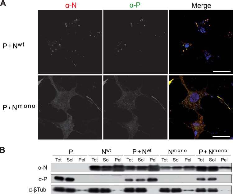
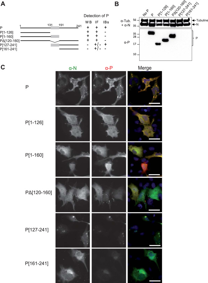
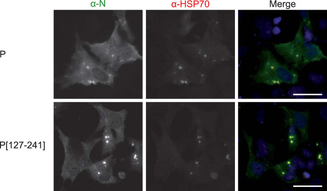
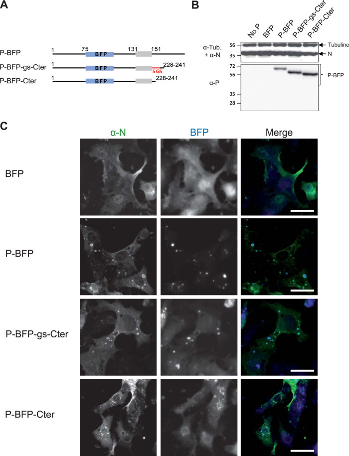
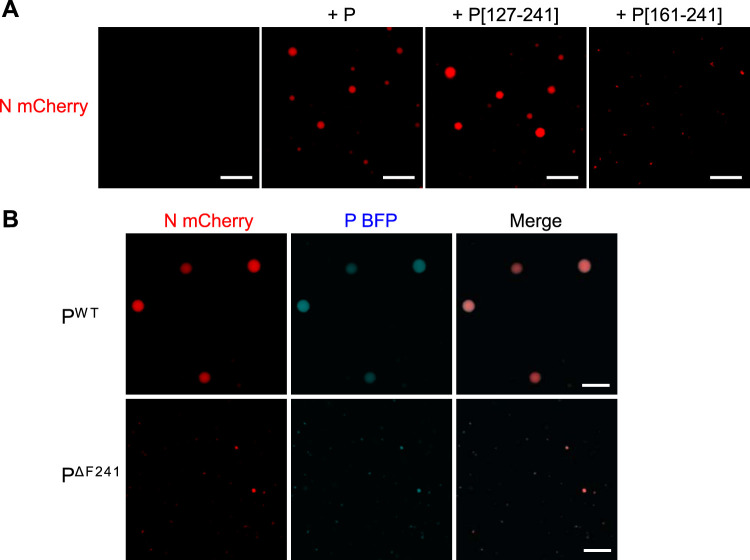
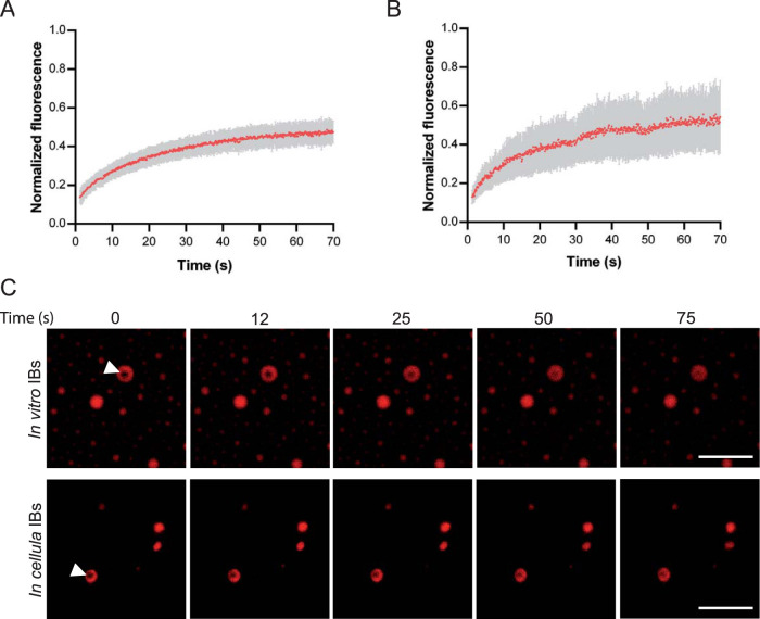
Infection of host cells by the respiratory syncytial virus (RSV) is characterized by the formation of spherical cytoplasmic inclusion bodies (IBs). These structures, which concentrate all the proteins of the polymerase complex as well as some cellular proteins, were initially considered aggresomes formed by viral dead-end products. However, recent studies revealed that IBs are viral factories where viral RNA synthesis, i.e., replication and transcription, occurs. The analysis of IBs by electron microscopy revealed that they are membrane-less structures, and accumulated data on their structure, organization, and kinetics of formation revealed that IBs share the characteristics of cellular organelles, such as P-bodies or stress granules, suggesting that their morphogenesis depends on a liquid-liquid phase separation mechanism. It was previously shown that expression of the RSV nucleoprotein N and phosphoprotein P of the polymerase complex is sufficient to induce the formation of pseudo-IBs. Here, using a series of truncated P proteins, we identified the domains of P required for IB formation and show that the oligomeric state of N, provided it can interact with RNA, is critical for their morphogenesis. We also show that pseudo-IBs can form in vitro when recombinant N and P proteins are mixed. Finally, using fluorescence recovery after photobleaching approaches, we reveal that in cellula and in vitro IBs are liquid organelles. Our results strongly support the liquid-liquid phase separation nature of IBs and pave the way for further characterization of their dynamics.IMPORTANCE Respiratory syncytial virus (RSV) is the leading cause of lower respiratory tract illness in infants, elderly, and immunocompromised people. No vaccine or efficient antiviral treatment is available against this virus. The replication and transcription steps of the viral genome are appealing mechanisms to target for the development of new antiviral strategies. These activities take place within cytoplasmic inclusion bodies (IBs) that assemble during infection. Although expression of both the viral nucleoprotein (N) and phosphoprotein (P) allows induction of the formation of these IBs, the mechanism sustaining their assembly remains poorly characterized. Here, we identified key elements of N and P required for the scaffolding of IBs and managed for the first time to reconstitute RSV pseudo-IBs in vitro by coincubating recombinant N and P proteins. Our results provide strong evidence that the biogenesis of RSV IBs occurs through liquid-liquid phase transition mediated by N-P interactions.

Keywords: RSV; inclusion bodies; liquid-liquid phase; nucleoprotein; phosphoprotein; protein-protein interactions.


Figures

FIG 1
FIG 1
Study of P-N interactions in cells by immunofluorescence and soluble-insoluble fractionation. (A) Observation of cellular localization of P and Nwt or Nmono in cells. N and P proteins were coexpressed in BSRT7/5 cells. The cells were then fixed 24 h posttransfection and labeled with anti-P (α-P) (green) and anti-N (α-N) (red) antibodies, and the distribution of viral proteins was observed by fluorescence microscopy. Nuclei were stained with Hoechst 33342. Bars, 10 μm. (B) BSRT7/5 cells expressing P, Nwt, or Nmono proteins or coexpressing P and Nwt or Nmono were lysed 24 h posttransfection, and the presence of P and N proteins in total (Tot), soluble (Sol), and insoluble (pellet [Pel]) fractions were analyzed by Western blotting.
FIG 2
FIG 2
Identification of the domains of P required for the formation of IBs in cells. (A) Schematic illustration of truncated P proteins. The oligomerization domain of P is represented in gray, and numbers indicate amino acid positions. For each deletion mutant, the detection of P by Western blotting (WB), immunolabeling (immunofluorescence [IF]), and within IBs, by the rabbit polyclonal antibody, is summarized to the right of the schematic. (B) Western blot analysis of the expression of N and P fragments in BRST/7 cells cotransfected with pN and pP (or fragments). Detection of tubulin was used as a control. (C) Cellular localization of Nwt and P fragments in cells. N and P proteins were coexpressed in BSRT7/5 cells. The cells were then fixed 24 h posttransfection and labeled with anti-N (green) and anti-P (red) antibodies, and the distribution of viral proteins was observed by fluorescence microscopy. Nuclei were stained with Hoechst 33342. Bars, 20 μm.
FIG 3
FIG 3
Analysis of the recruitment of HSP70 to IBs. Cells were cotransfected with N and P or P[127-241], fixed 24 h posttransfection, and labeled with anti-N (green) and anti-HSP70 (red) antibodies before observation by fluorescence microscopy. Nuclei were stained with Hoechst 33342. Bars, 20 μm.
FIG 4
FIG 4
Study of the role of the intrinsically disordered domain [160-227] of P for IB formation. (A) Schematic illustration of the full-length and truncated P-BFP proteins. The oligomerization domain of P is represented in gray. The BFP is shown in blue, and the sequence of five Gly-Ser repetition is shown in red. Numbers indicate amino acid positions. (B) Western blot analysis of the expression of N and P-BFP proteins in BRST/7 cells cotransfected with pN- and pP-BFP-derived constructs. Detection of tubulin was used as a control. (C) Cellular localization of N and P-BFP proteins in cells. N and P-BFP proteins were coexpressed in BSRT7/5 cells. The cells were then fixed 24h posttransfection and labeled with anti-N (green) antibodies, and the distribution of viral proteins was observed by fluorescence microscopy. Bars, 20 μm.
FIG 5
FIG 5
Reconstitution of pseudo-IBs in vitro. Recombinant N and P proteins were coincubated, and the analysis of phase separation was assessed using fluorescence microscopy. (A) Coincubation of mCherry-N (3 μM) and P-BFP (14 μM) in the presence of increasing concentratiosn of Ficoll. (B) Coincubation of mCherry-N (3 μM) and increasing concentration of P-BFP (from 3 μM to 14 μM) in the presence of 15% Ficoll. The ratio of mCherry N to P-BFP is indicated. (C) Analysis of purified N, mCherry-N, P, and P-BFP recombinant proteins by SDS-PAGE and Coomassie blue staining. (D) Coincubation of P-BPF and Nwt or mCherry-N and Pwt in the presence of 15% Ficoll. Bars, 10 μm.
FIG 6
FIG 6
Validation of the minimal domains of P required for pseudo-IB formation in vitro. Recombinant N and P proteins were coincubated, and the analysis of phase separation was assessed using fluorescence microscopy. (A) Coincubation of mCherry-N (3 μM) with recombinant P or the fragments P[127-241] and P[161-241] (35 μM), 15% Ficoll; (B) coincubation of mCherry-N with recombinant Pwt-BFP or P ΔF241-BFP. Bars, 10 μm.
FIG 7
FIG 7
Study of IB viscosity. FRAP analysis of mCherry-N fluorescence in IBs. (A and B) Spontaneous redistribution of mCherry-N fluorescence after photobleaching on in vitro (A) and cellular (B) IBs was recorded, corrected (background and bleaching during postbleach imaging) and normalized to the average of the prebleach signal. Data are from 20 FRAP events recorded in two independent experiments. The mean of each experimental condition is shown with error bars representing standard deviations (SD). (C) Time-lapse images of FRAP on in vitro IBs (top panels), and on pseudo-IBs formed in cells transfected with pmCherry-N and pP for 24 h (bottom panels). Bars, 10 μm.

References

    1. Coultas JA, Smyth R, Openshaw PJ. 2019. Respiratory syncytial virus (RSV): a scourge from infancy to old age. Thorax 74:986–993. doi:10.1136/thoraxjnl-2018-212212. - DOI - PubMed
    1. Wang D, Cummins C, Bayliss S, Sandercock J, Burls A. 2008. Immunoprophylaxis against respiratory syncytial virus (RSV) with palivizumab in children: a systematic review and economic evaluation. Health Technol Assess 12:iii, ix–x, 1–86. doi:10.3310/hta12360. - DOI - PubMed
    1. Afonso CL, Amarasinghe GK, Bányai K, Bào Y, Basler CF, Bavari S, Bejerman N, Blasdell KR, Briand F-X, Briese T, Bukreyev A, Calisher CH, Chandran K, Chéng J, Clawson AN, Collins PL, Dietzgen RG, Dolnik O, Domier LL, Dürrwald R, Dye JM, Easton AJ, Ebihara H, Farkas SL, Freitas-Astúa J, Formenty P, Fouchier RAM, Fù Y, Ghedin E, Goodin MM, Hewson R, Horie M, Hyndman TH, Jiāng D, Kitajima EW, Kobinger GP, Kondo H, Kurath G, Lamb RA, Lenardon S, Leroy EM, Li C-X, Lin X-D, Liú L, Longdon B, Marton S, Maisner A, Mühlberger E, Netesov SV, Nowotny N, et al. 2016. Taxonomy of the order Mononegavirales: update 2016. Arch Virol 161:2351–2360. doi:10.1007/s00705-016-2880-1. - DOI - PMC - PubMed
    1. Bakker SE, Duquerroy S, Galloux M, Loney C, Conner E, Eléouët J-F, Rey FA, Bhella D. 2013. The respiratory syncytial virus nucleoprotein-RNA complex forms a left-handed helical nucleocapsid. J Gen Virol 94:1734–1738. doi:10.1099/vir.0.053025-0. - DOI - PMC - PubMed
    1. Collins PL, Melero JA. 2011. Progress in understanding and controlling respiratory syncytial virus: still crazy after all these years. Virus Res 162:80–99. doi:10.1016/j.virusres.2011.09.020. - DOI - PMC - PubMed

Publication types

LinkOut - more resources