. 2020 Sep 9:2020:9758156.
doi: 10.1155/2020/9758156. eCollection 2020.

Extract of Pogostemon cablin Possesses Potent Anticancer Activity against Colorectal Cancer Cells In Vitro and In Vivo

Affiliations

Extract of Pogostemon cablin Possesses Potent Anticancer Activity against Colorectal Cancer Cells In Vitro and In Vivo

Ju-Huei Chien et al. Evid Based Complement Alternat Med. .

Abstract

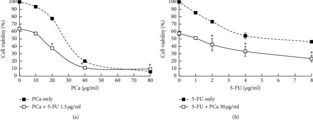
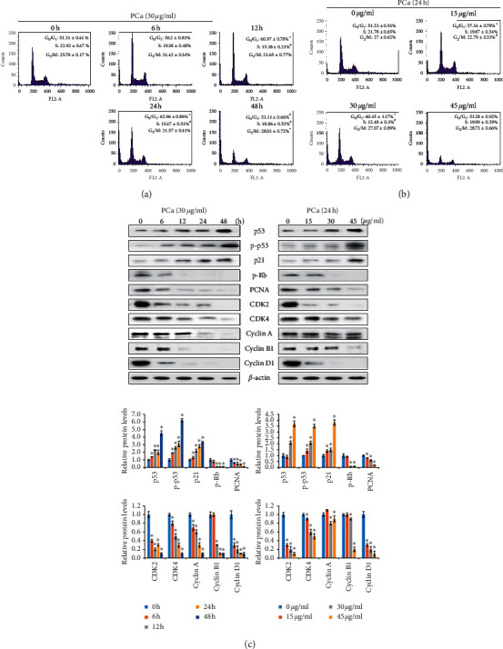
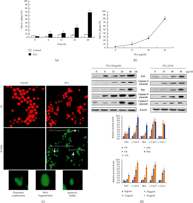
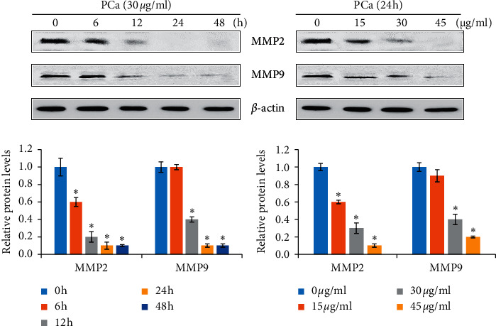
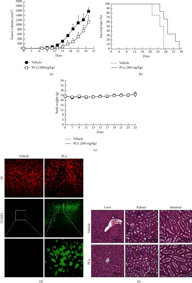
Pogostemon cablin (PCa), an herb used in traditional Chinese medicine, is routinely used in the amelioration of different types of gastrointestinal discomfort. However, the mechanisms underlying the cancer suppression activity of PCa in colorectal cancer (CRC) cells have yet to be clarified. The aim of this study was to investigate the anticancer effects of PCa, specifically the induction of apoptosis in CRC cells. The growth inhibition curve of CRC cells following exposure to PCa was detected by an MTT assay. Moreover, PCa combined with 5-FU revealed a synergic effect of decreased cell viability. PCa inhibited cell proliferation and induced cell cycle arrest at the G0/G1 phase and cell apoptosis through regulation of associated protein expression. An in vivo study showed that PCa suppressed the growth of CRC via induction of cell apoptosis with no significant change in body weight or organ histology. Our results demonstrated that PCa inhibits the growth of CRC cells and induces apoptosis in vitro and in vivo, which suggests the potential applicability of PCa as an anticancer agent.


Conflict of interest statement

The authors declare no conflicts of interest regarding the publication of this paper.

Figures

Figure 1
Figure 1
Effect of PCa on cell survival and proliferation of CRC cells. Cell viability was determined by using an MTT assay after treatment for 24, 48, or 72 h with increasing concentrations of PCa. (a) HT-29, (b) CT26, (c) SVEC, and (d) MDCK cells. The percentage of cell viability was normalized to the control (100%). The results are expressed as mean ± SD.
Figure 2
Figure 2
PCa and 5-FU synergistically inhibited the growth of HT-29 cells in vitro. HT-29 cells were treated with (a) PCa (0, 10, 20, 40, or 80 μg/ml) and/or 5-FU (1.5 μg/ml), (b) 5-FU (0, 1 2, 4, or 8 μg/ml) and/or PCa (30 μg/ml) for 48 h, and then we measured cell viability by using an MTT assay. PCa and 5-FU revealed a synergistic effect (CI value < 1). Compared with PCa or 5-FU only vs. the combination group (P < 0.05).
Figure 3
Figure 3
PCa induced G0/G1 phase cell cycle arrest by decreasing CDK4 and cyclin D1 expression. Cell cycle distribution (G0/G1, S, G2/M phases) of HT-29 cells after treatment with 30 μg/ml PCa for 0–48 h (a); 15, 30, or 45 μg/ml PCa for 24 h (b) was detected and analyzed by flow cytometry and FlowJo software. The results are shown as mean ± SD. P < 0.05 versus control group with a significant increase. #P < 0.05 versus control group with a significant decrease. (c) Western blot showing the upregulation of the proteins p53 and p21, whereas downregulation of the proteins CDK4 and cyclin D1 was observed in HT-29 cells.
Figure 4
Figure 4
PCa triggered cell apoptosis through extrinsic and intrinsic pathways in HT-29 cells. (a, b) HT-29 cells were treated with PCa and analyzed for SubG1 phase by flow cytometry. Data are expressed as mean ± SD. P < 0.05 versus control group with a significant increase. (c) The typical morphology of cell apoptosis, such as chromatin condensation, DNA fragmentation, and apoptosis body, was observed after PCa treatment for 48 h by using TUNEL assays. (d) Determination of the apoptosis pathway by Western blots of PCa-treated cells.
Figure 5
Figure 5
The molecules involved in metastasis were affected by PCa treatment of HT-29 cells. Cells were treated with 30 μg/ml PCa for 0, 6, 12, 24, or 48 h; 15, 30, or 45 μg/ml PCa for 24 h, and the protein expression of MMP2 and MMP9 was analyzed by Western blotting. Protein bands were normalized using the corresponding β-actin to calculate the band density. MMP: matrix metallopeptidase.
Figure 6
Figure 6
PCa suppressed tumor growth via induction of cell apoptosis in vivo. Mice were subcutaneously implanted with CT26 cells and treated (s.c.) with 200 mg/kg PCa once every two days for 40 days. Tumor volume (a), survival (b), and body weight (c) were recorded once every two days, and mice were sacrificed when the volume of the tumor exceeded 1500 mm3. Results are expressed as mean ± SD. P < 0.05 versus the vehicle group. (d) Cell apoptosis was determined through tissue TUNEL assays. (e) HE staining of the organs, including liver, kidney, and intestine, was used for assessing toxicity.

References

    1. Coleman M. P., Quaresma M., Berrino F., et al. Cancer survival in five continents: a worldwide population-based study (CONCORD) The Lancet Oncology. 2008;9(8):730–756. doi: 10.1016/s1470-2045(08)70179-7. - DOI - PubMed
    1. Ferlay J., Colombet M., Soerjomataram I., et al. Estimating the global cancer incidence and mortality in 2018: GLOBOCAN sources and methods. International Journal of Cancer. 2019;144(8):1941–1953. doi: 10.1002/ijc.31937. - DOI - PubMed
    1. Labianca R., Beretta G. D., Kildani B., et al. Colon cancer. Critical Reviews in Oncology/Hematology. 2010;74(2):106–133. doi: 10.1016/j.critrevonc.2010.01.010. - DOI - PubMed
    1. De Dosso S., Sessa C., Saletti P. Adjuvant therapy for colon cancer: present and perspectives. Cancer Treatment Reviews. 2009;35(2):160–166. doi: 10.1016/j.ctrv.2008.10.001. - DOI - PubMed
    1. Kopetz S., Freitas D., Calabrich A. F., Hoff P. M. Adjuvant chemotherapy for stage II colon cancer. Oncology (Williston Park) 2008;22(3):260–270. - PubMed

LinkOut - more resources