. 2020 May-Jun;11(3):289-299.
doi: 10.32598/bcn.11.2.1189.1. Epub 2020 May 1.

Protective Effect of Curcumin on Hippocampal and Behavior Changes in Rats Exposed to Fluoride During Pre- and Post-natal Period

Affiliations

Protective Effect of Curcumin on Hippocampal and Behavior Changes in Rats Exposed to Fluoride During Pre- and Post-natal Period

Nagapuri Kiran Kumar et al. Basic Clin Neurosci. 2020 May-Jun.

Abstract

Introduction: Curcumin, a yellow-pigment, found in the popular Indian spice turmeric (Curcuma longa), poses pharmaceutical applications due to its anti-inflammatory, antioxidant, and chemoprotective properties. Excessive fluoride causes fluorosis leading to neurodegeneration and associated behavioral deficits, particularly in children. This study aimed at investigating the neuroprotective ability of curcumin on sodium fluoride (NaF)-related alterations of acetylcholine, catecholamines, histological changes in hippocampus and behavior of rats exposed to NaF during pre- and post-natal period.

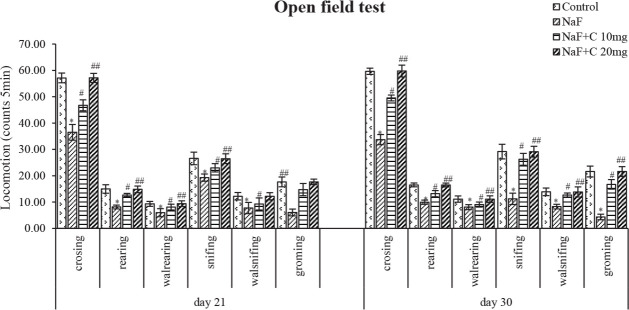
Methods: Pregnant albino Wistar rats were chosen and divided into four groups. The experimental period lasted 53 days (i.e. the gestational period of 23 days and post-gestational period of 30 days), at which the control group received normal tap water, the experimental group received NaF (20 ppm/kg bw) through drinking water, and the protective groups received curcumin (10 mg and 20mg/kg bw) by gavage and NaF (20 ppm/kg bw) through drinking water. Behavioral study (open field test) was done using postnatal pups aged 21 and 30 days. The brains of postnatal pups aged 1, 7, 14, 21, and 30 days were collected and used for biochemical analysis and those of pups aged 14, 21, and 30 days were used for histopathological analysis.

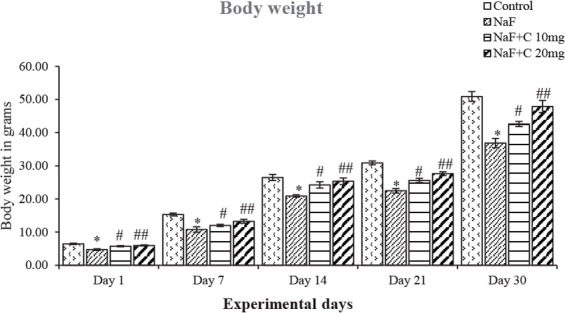
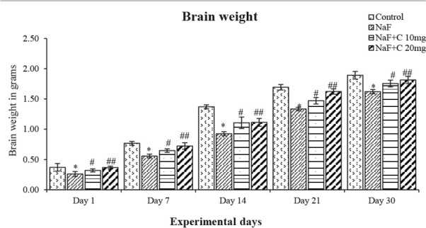
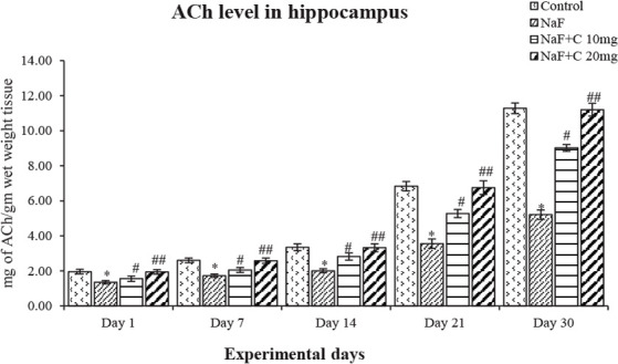
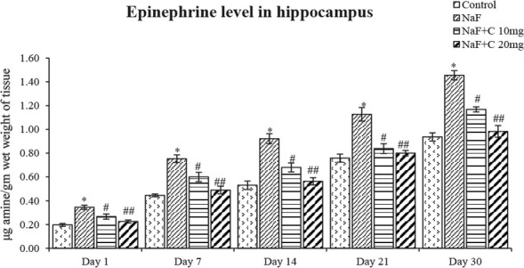
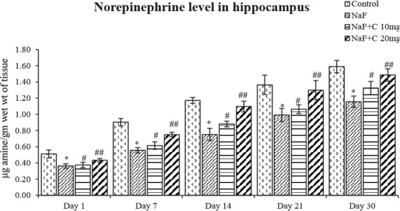
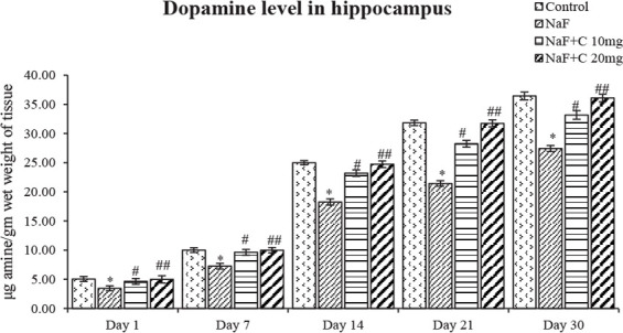
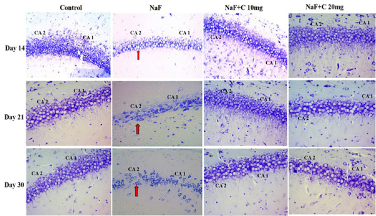
Results: NaF-exposed rats showed a significant (P<0.05) decrease in body weight, brain weight, and behavioral activities, which were significantly reversed with curcumin treatment. The levels of epinephrine significantly (P<0.05) increased, whereas norepinephrine, dopamine and acetylcholine levels declined in NaF-treated group compared with the control group, which were significantly (P<0.05) reversed after treatment by curcumin (10 mg/kg bw and 20 mg/kg bw) along with NaF. The histological alterations, including shrinkage of neurons and nissal substances were observed in the hippocampus of NaF-treated pups that the control pups, whereas co-treatment with curcumin and NaF showed ameliorative effects and controlled the histological alterations.

Conclusion: The results showed the neuroprotective effect of curcumin on behavior, neurotransmitter levels, and histological changes in the hippocampus against NaF-induced neurotoxicity in developing rat pups.

Keywords: Behavior; Curcumin; Hippocampus; Neurotransmitters; Sodium fluoride.


Conflict of interest statement

Conflict of interest The authors declared no conflict of interest.

Figures

Figure 1.
Figure 1.
Effect of curcumin on body weight of the rats treated with sodium fluoride (NaF). * P<0.05 vs. the control group. #P<0.05 and ##P<0.05 vs. the NaF-treated group. Values are expressed as Mean±SD and the results are reported in grams (g); (n=6).
Figure 2.
Figure 2.
Effect of curcumin on brain weight of the rats treated with sodium fluoride (NaF). *P<0.05 vs. the control group. #P<0.05 and ##P<0.05 vs. NaF-treated group. Values are expressed as Mean±SD and the results are reported in grams (g); (n=6).
Figure 3.
Figure 3.
Effect of curcumin on Open field test results in rats treated with sodium fluoride. *P<0.05 vs. the control group. #P<0.05 and ##P<0.05 vs. sodium fluoride (NaF)-treated group. Values are expressed as Mean±SEM and the results are reported as the number of entries (counts/5 min); (n=6)
Figure 4.
Figure 4.
Acetylcholine levels in the developing hippocampus of the rats treated with sodium fluoride (NaF) and curcumin. * P<0.05 vs. the control group; #P<0.05 and ## P<0.05 vs. NaF-treated group. Values are expressed as Mean±SEM and the results are reported in mg of ACh/gm wet weight tissue; (n=6).
Figure 5.
Figure 5.
Epinephrine levels in the developing hippocampus of the rats treated with sodium fluoride (NaF) and curcumin * P<0.05 vs. the control group. #P<0.05 and ##P<0.05 vs. the NaF-treated group. Values are expressed as Mean±SEM and the results are reported in μg amine/gm wet weight of tissue; (n=6).
Figure 6.
Figure 6.
Norepinephrine levels in the developing hippocampus of rats treated with sodium fluoride (NaF) and curcumin. *P<0.05 vs. the control group; #P<0.05 and ##P<0.05 vs. the NaF-treated group. Values are expressed as Mean±SEM and the results are reported in μg amine/gm wet wt of tissue; (n=6).
Figure 7.
Figure 7.
Dopamine levels in developing hippocampus of the rats treated with sodium fluoride (NaF) and curcumin. * P<0.05 vs. the control group; # P<0.05 and ## P<0.05 vs. the NaF-treated group. Values are expressed as Mean±SEM and the results are reported in μg amine/gm wet weight of tissue; (n=6).
Figure 8.
Figure 8.
Histopathological studies in the hippocampal CA1 and CA2 regions stained by cresyl violet in the control group and groups treated with sodium fluoride (NaF), NaF+curcumin (10 mg), and NaF+curcumin (20 mg) on postnatal days 14, 21, and 30. The neurons were arranged closely, had a circular shape, and the nuclear membrane was clear in the hippocampal CA1 and CA2 regions of the control group and groups treated with NaF+curcumin (10 mg) and NaF+curcumin (20 mg) groups. Arrow marks show the neurons with irregular and shrunken shapes, and the nuclear membrane was indistinct in the CA1 and CA2 regions in the NaF-treated group (Magnification 40X).

Similar articles

Cited by

References

    1. Arora V. A., Kuhad V., Tiwari K. C. (2011). Curcumin ameliorates reserpine-induced pain-depression dyad: Behavioural, biochemical, neurochemical and molecular evidences. Psychoneuroendocrinology, 36(10), 1570–81. [DOI:10.1016/j.psyneuen.2011.04.012] [PMID ] - DOI - PubMed
    1. Bhatnagar M., Rao P., Saxena A., Bhatnagar R., Meena P., Barbar S., Chouhan A., et al. (2006). Biochemical changes in brain and other tissues of young adult female mice from fluoride in their drinking water. Fluoride, 39(4), 280–4.
    1. Brown L., Buckingham S., Jones A. (2009). Nicotinic Acetylcholine Receptor Signaling: Roles in Alzheimer’s disease and Amyloid Neuroprotection. Pharmacological Review, 61(1), 39–61. [DOI:10.1124/pr.108.000562] [PMID ] [PMCID ] - DOI - PMC - PubMed
    1. Chandrashakar R. N., Reddy K.P. (2016). Protective effect of resveratrol against neuronal damage through oxidative stress in cerebral hemisphere of aluminum and fluoride treated rats. Interdisciplinary Toxicology, 9(2), 78–82. [DOI:10.1515/intox-2016-0009] [PMID ] [PMCID ] - DOI - PMC - PubMed
    1. Chhavi S., Pooja S., Piyu S., Neha J., Maheep B. (2014). Curcumin attenuates neurotoxicity induced by fluoride: An in vivoevidence. Pharmacoghosy Magazine, 10(37), 61–5. [DOI:10.4103/0973-1296.126663] [PMID ] [PMCID ] - DOI - PMC - PubMed

LinkOut - more resources