. 2020 Sep 10:6:84.
doi: 10.1038/s41420-020-00318-7. eCollection 2020.

MFG-E8 accelerates wound healing in diabetes by regulating "NLRP3 inflammasome-neutrophil extracellular traps" axis

Affiliations

MFG-E8 accelerates wound healing in diabetes by regulating "NLRP3 inflammasome-neutrophil extracellular traps" axis

Wei Huang et al. Cell Death Discov. .

Abstract

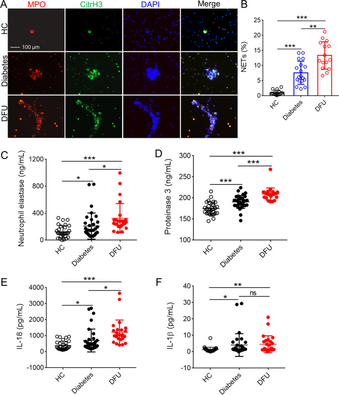
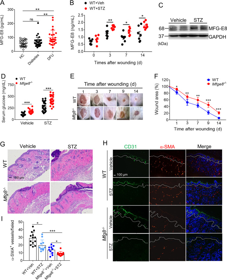
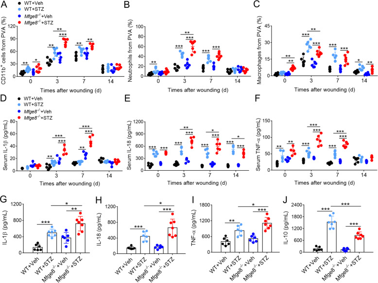
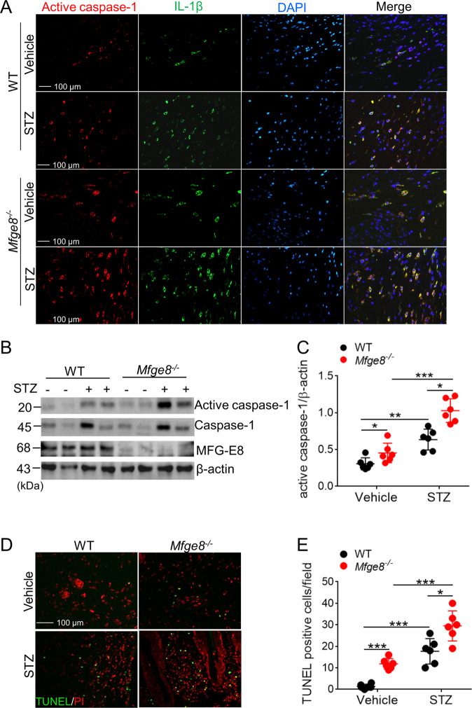
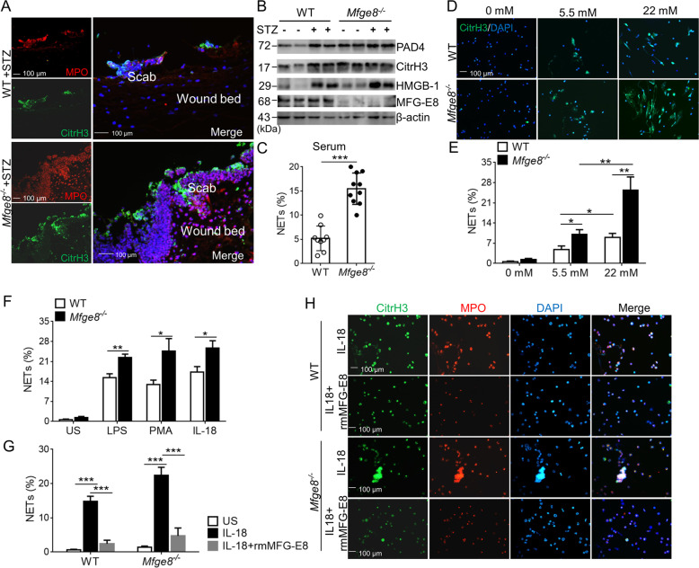
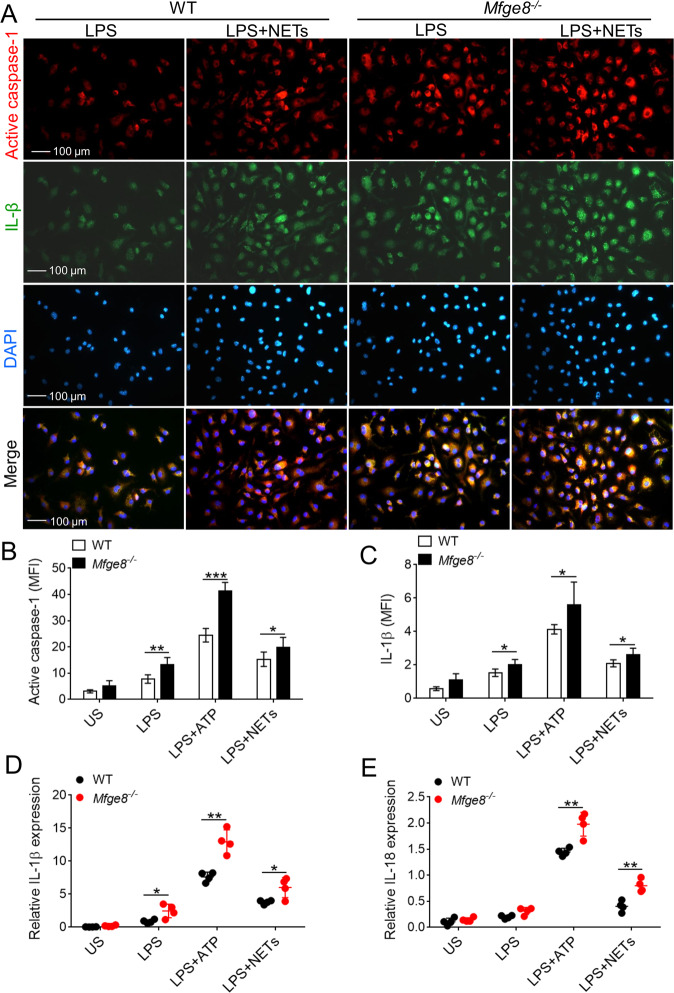
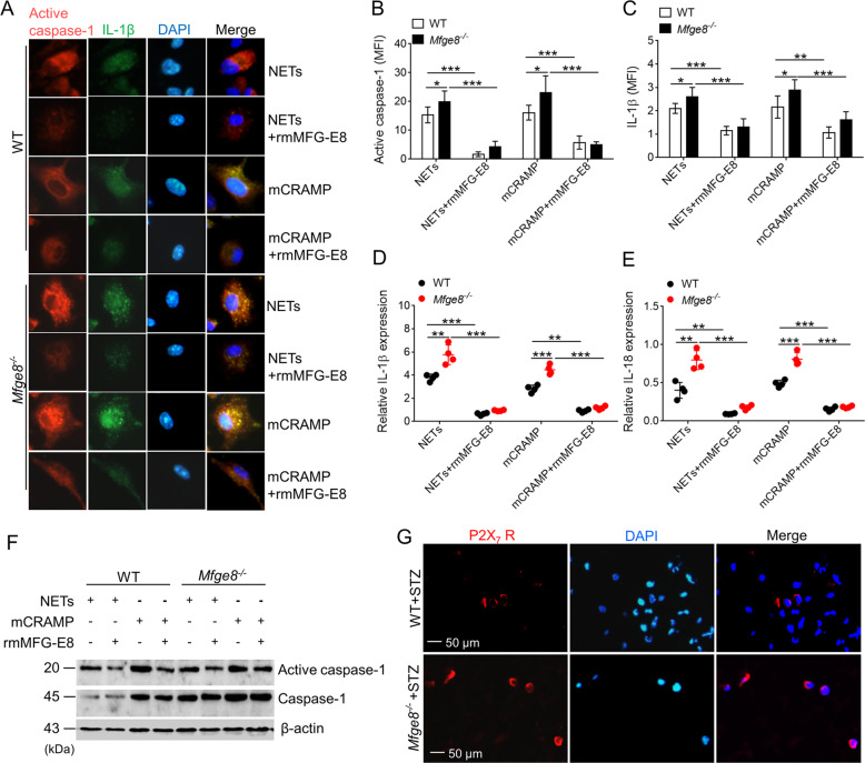
Sustained activation of NLRP3 inflammasome and release of neutrophil extracellular traps (NETs) impair wound healing of diabetic foot ulcers (DFUs). Our previous study reported that milk fat globule epidermal growth factor VIII (MFG-E8) attenuates tissue damage in systemic lupus erythematosus. However, the functional effect of MFG-E8 on "NLRP3 inflammasome-NETs" inflammatory loop in wound healing of diabetes is not completely elucidated. In this study, neutrophils from DFU patients are susceptible to undergo NETosis, releasing more NETs. The circulating levels of NET components neutrophil elastase and proteinase 3 and inflammatory cytokines IL-1β and IL-18 were significantly elevated in DFU patients compared with healthy controls or diabetic patients, in spite of higher levels of MFG-E8 in DFU patients. In Mfge8-/- diabetic mice, skin wound displayed exaggerated inflammatory response, including leukocyte infiltration, excessive activation of NLRP3 inflammasome (release of higher IL-1β, IL-18, and TNF-α), largely lodged NETs, resulting in poor angiogenesis and wound closure. When stimulated with high-dose glucose or IL-18, MFG-E8-deficient neutrophils release more NETs than WT neutrophils. After administration of recombinant MFG-E8, IL-18-primed NETosis of WT or Mfge8-/- neutrophils was significantly inhibited. Furthermore, NET and mCRAMP (component of NETs, the murine equivalent of cathelicidin LL-37 in human)-mediated activation of NLRP3 inflammasome and production of IL-1β/IL-18 were significantly elevated in Mfge8-/- macrophages compared with WT macrophages, which were also significantly dampened by the administration of rmMFG-E8. Therefore, our study demonstrated that as inhibitor of the "NLRP3 inflammasome-NETs" inflammatory loop, exogenous rMFG-E8 improves angiogenesis and accelerates wound healing, highlighting possible therapeutic potential for DFUs.

Keywords: Cell death and immune response; Diabetes complications; Inflammasome.


Conflict of interest statement

Conflict of interestThe authors declare that they have no conflict of interest.

Figures

Fig. 1
Fig. 1. The activation of NLRP3 inflammasome and NETosis in patients with DFUs.
a Neutrophils were purified from peripheral blood of HCs (n = 10), diabetes (n = 12), and DFU patients (n = 12) under spontaneous NETosis without FBS cultivation; and visualization of NETs with citrullinated histone H3 (CitrH3), MPO, and DNA. Three colors were merged by software Image J (NIH), original magnification ×200. b NET% was calculated in neutrophils from HCs, diabetic, and DFU patients (NET% = positive enlarged nuclei/total neutrophils per field, and the average of NET release ratio was evaluated >500 cells in more than five fields per sample were counted. cf Serum neutrophil elstase c, proteinase 3 d, IL-18 e, and IL-1β f concentration in HCs (n = 30), diabetes (n = 33), and DFU patients (n = 25) were detected with ELISA. For all experiments, data are presented as mean ± SEM, *P < 0.05, **P < 0.01, ***P < 0.001, ns = not significant.
Fig. 2
Fig. 2. MFG-E8 deficiency impairs wound closure and wound angiogenesis.
a The serum levels of MF-E8 in HCs (n = 30), diabetes (n = 33), and DFU patients (n = 25) were detected with ELISA. b The serum levels of MFG-E8 in normal or diabetic mice (n = 6) post-wounding for 0, 3, 7, 14 days were determined by ELISA. c Western blot analysis of MFG-E8 expression in skin tissues of normal or diabetic mice after wound for 3 days, GAPDH as a loading control. d The serum fed glucose levels of WT (n = 24) or Mfge8−/− mice (n = 28) when treated with vehicle or STZ for 28 days were measured. e Representative digital imaging of wounds from age-matched diabetic WT or Mfge8−/− mice postwounding up to day 14. f Wound closures kinetics of diabetic WT and Mfge8−/− mice (n = 6). g Representative images of H&E-stained skin tissues from WT or Mfge8−/− mice injected with vehicle or STZ after wound for 3 days. h The amount of CD31+ epithelial cells (ECs) and α-smooth muscle actin (α-SMA+) pericytes in wound skins of WT or Mfge8−/− mice treated with vehicle or STZ at day 14 after wound. i Quantification of α-SMA+ vessels in five random microscopic fields in n = 6 mice per group was performed. For all experiments, data are presented as mean ± SEM, *P < 0.05, **P < 0.01, ***P < 0.001.
Fig. 3
Fig. 3. MFG-E8 deficiency enhances inflammatory response at the wound sites of diabetic mice.
ac Statistical analysis of total CD11b+ cells a, CD11b+ Ly6G+ neutrophils b, and CD11b+ F4/80+ macrophages c in PVA wound sites of WT or Mfge8−/− (n = 6) mice treated with vehicle or STZ post-wounding at day 0, 3, 7, and 14. df Inflammatory cytokine profiles response to wound. The serum levels of IL-1β d, IL-18 e, and TNF-α f from WT or Mfge8−/− mice (n = 6) treated with vehicle or STZ post-wounding at day 0, 3, 7, and 14 were measured with ELISA. gj Cytokine profiles in wound site. The concentration of IL-1β g, IL-18 h, TNF-α i, and IL-10 j in PVA from wound skins of WT or Mfge8−/− mice (n = 6) treated with vehicle or STZ post-wounding at day 3 were determined with ELISA. For all experiments, data are presented as mean ± SEM, *P < 0.05, **P < 0.01, ***P < 0.001.
Fig. 4
Fig. 4. The activation of NLRP3 inflammasome was aggravated in MFG-E8-deficient mice.
a The infiltrated active caspase-1+ and IL-1β+ macrophages in dermis of wound skins from WT or Mfge8−/− mice (n = 6) treated with vehicle or STZ, post-wounding at day 3, were determined with immunofluorescence. b Quantification of the expression of active caspase-1, caspase-1, and MFG-E8 in wound skin tissue from WT or Mfge8−/− mice (n = 6) treated with vehicle or STZ at day 3 post-wounding, β-actin as loading control. c Calculation of the ratio of active caspase-1/caspase-1 in wound skin tissues from each group mice. d Infiltration of death cells into wound skin tissues of WT or Mfge8−/− mice (n = 6) post-wounding at day 3 was identified by TUNEL. Representative images were shown, original magnification ×200. e The average TUNEL-positive cells were counted at more than five random microscopic fields. For all experiments, data are presented as mean ± SEM, *P < 0.05, **P < 0.01, ***P < 0.001.
Fig. 5
Fig. 5. MFG-E8 attenuates NETosis in diabetic wound mice.
a Immunofluorescence images of NETs in the wound bed beneath the scab from diabetic WT or Mfge8−/− mice (n = 6) after wound for 3 days, visualized with DNA, CitrH3, and MPO. Three color were merged by software Image J (NIH). b Western blot of the wound tissues collected post-wounding at day 3 from WT or Mfge8−/− mice (n = 6) treated with vehicle or STZ, to detect the expression of PAD4, CitrH3, HMGB-1, and MFG-E8, β-actin as loading control. c WT neutrophils primed NETosis, stimulated with the serum from diabetic WT or Mfge8−/− mice (n = 6) post-wounding at day 3, and the percentage of NET formation were calculated. d Representative immunofluorescence images of NETs from isolated neutrophils of WT or Mfge8−/− mice (n = 6) exposed to different glucose concentrations in vitro. e Percentage of produced NETs in neutrophils from WT or Mfge8−/− mice (n = 5) after exposure with normal or high glucose were displayed. f The in vitro NET release of WT or Mfge8−/− neutrophils (n = 5) was detected after stimulation with PMA, LPS, or IL-18. US, unstimulated. g After treatment with 500 ng/mL rmMFG-E8, the NET% were calculated in WT or Mfge8−/− neutrophils (n = 5) stimulated with IL-18. h The representative images of NET release from isolated WT or Mfge8−/− neutrophils (n = 5) induced by IL-18. For all experiments, data are presented as mean ± SEM, *P < 0.05, **P < 0.01, ***P < 0.001.
Fig. 6
Fig. 6. The activation of NLRP3 inflammasome and release of IL-1β and IL-18 were elevated in MFG-E8-deficient macrophages.
a Representative immunofluorescence images of LPS-primed BMDMs from WT or Mfge8−/− stained with active caspase-1 and IL-1β after stimulated with 10 μg/mL NETs. The active caspase-1+ and IL-1β+ BMDMs in five random microscopic fields in n = 4 mice per group was performed, original magnification ×400. bc Quantified the mean fluorescence density (MFI) of active caspase-1 b and IL-1β c in LPS-primed BMDMs from WT or Mfge8−/− mice (n = 4) after treatment with NETs or ATP. de Real-time PCR analysis of IL-1β d and IL-18 e mRNA levels in LPS-primed and ATP or NETs-stimulated WT or Mfge8−/− BMDMs (from n = 4 mice respectively). For all experiments, data are presented as mean ± SEM, *P < 0.05, **P < 0.01, ***P < 0.001.
Fig. 7
Fig. 7. MFG-E8 inhibits the NETs-induced activation of NLRP3 inflammasome.
a Representative immunofluorescence images of active caspase-1 and IL-1β in LPS-primed BMDMs from WT or Mfge8−/− mice (n = 4) treated with 2 μg/mL mCRAMP (LL-37) or 10 μg/mL NETs, and then administrated with or without rmFMG-E8. The active caspase-1+ and IL-1β+ BMDMs in five random microscopic fields in n = 4 mice per group was performed. bc After administration with or without rmMFG-E8, quantified the mean fluorescence density (MFI) of active caspase-1 b and IL-1β c in LPS-primed BMDMs from WT or Mfge8−/− mice exposed to NETs or mCRAMP. de Quantification of IL-1β d and IL-18 e mRNA levels in LPS-primed BMDMs from WT or Mfge8−/− mice (n = 4) after stimulation with NETs or mCRAMP when addition with or without rmMFG-E8. f Exposure to NETs or mCRAMP, western blot analysis of the active caspase-1 and caspase-1 expression in BMDMs from WT or Mfge8−/− mice after treatment with or without rmMFG-E8. g The P2X7 receptor (P2X7 R+) macrophages in wound dermis from diabetic WT and Mfge8−/− mice were determined by immunofluorescence; more than five random microscopic fields in n = 6 mice per group was performed. For all experiments, data are presented as mean ± SEM, *P < 0.05, **P < 0.01, ***P < 0.001.

References

    1. Armstrong DG, Boulton AM, Bus SA. Diabetic foot ulcers and their recurrence. N. Engl. J. Med. 2017;376:2367–2375. doi: 10.1056/NEJMra1615439. - DOI - PubMed
    1. Sollberger G, Tilley DO, Zychlinsky A. Neutrophil extracellular traps: the biology of chromatin externalization. Dev. Cell. 2018;44:542–553. doi: 10.1016/j.devcel.2018.01.019. - DOI - PubMed
    1. Wong SL, et al. Diabetes primes neutrophils to undergo NETosis, which impairs wound healing. Nat. Med. 2015;21:815–817. doi: 10.1038/nm.3887. - DOI - PMC - PubMed
    1. Fadini GP, et al. NETosis delays diabetic wound healing in mice and humans. Diabetes. 2016;65:1061–1071. doi: 10.2337/db15-0863. - DOI - PubMed
    1. Menegazzo L, et al. NETosis is induced by high glucose and associated with type 2 diabetes. Acta Diabetol. 2015;52:497–503. doi: 10.1007/s00592-014-0676-x. - DOI - PubMed