

Bilateral Sesamoiditis as First Manifestation of Gout
- PMID: 32963862
- PMCID: PMC7499288
- DOI: 10.1155/2020/8890549
Bilateral Sesamoiditis as First Manifestation of Gout
Abstract
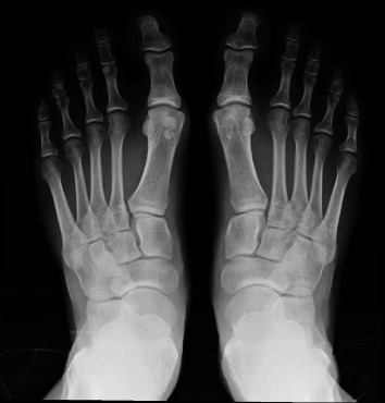
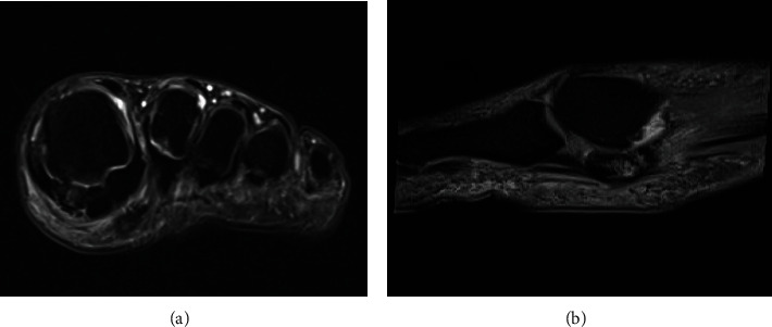
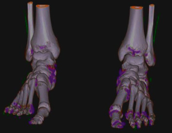
Sesamoiditis secondary to gout is an extremely rare condition with few case reports in the literature. It is an important differential diagnosis because the treatment depends on targeted therapy, unlike the main causes of sesamoiditis that often involves immobilization with special orthoses and prescription of anti-inflammatory drugs. We report here a case of a 38-year-old male, athlete, with bipartite medial sesamoid, who had insidious pain in the base of the left hallux. Laboratory tests showed no alterations, and imaging examinations demonstrated sesamoiditis with suspicion of stress fracture. The patient was initially prescribed an immobilization boot and analgesic and anti-inflammatory drugs, but he did not respond to the measures taken. After the onset of the same condition in the contralateral foot and getting the same imaging findings, we began an investigation of systemic disease, focusing on gout, because of a positive family history, which was confirmed by dual-energy computed tomography.
Copyright © 2020 Daniel de Oliveira Beraldo et al.
Conflict of interest statement
There are no conflicts of interest and source of funding.
Figures
References
-
- Perlman P. First metatarsal sesamoid pain. Australian Podiatrist. 1994;2:18–23.
Publication types
LinkOut - more resources
Full Text Sources
