. 2020 Aug;10(8):1534-1548.
doi: 10.1016/j.apsb.2020.02.002. Epub 2020 Feb 20.

Doxorubicin-loaded bacterial outer-membrane vesicles exert enhanced anti-tumor efficacy in non-small-cell lung cancer

Affiliations

Doxorubicin-loaded bacterial outer-membrane vesicles exert enhanced anti-tumor efficacy in non-small-cell lung cancer

Kudelaidi Kuerban et al. Acta Pharm Sin B. 2020 Aug.

Abstract

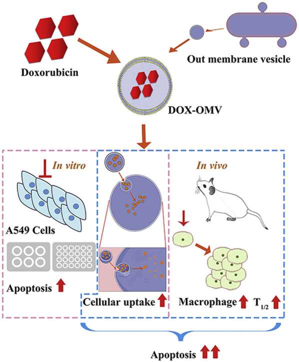
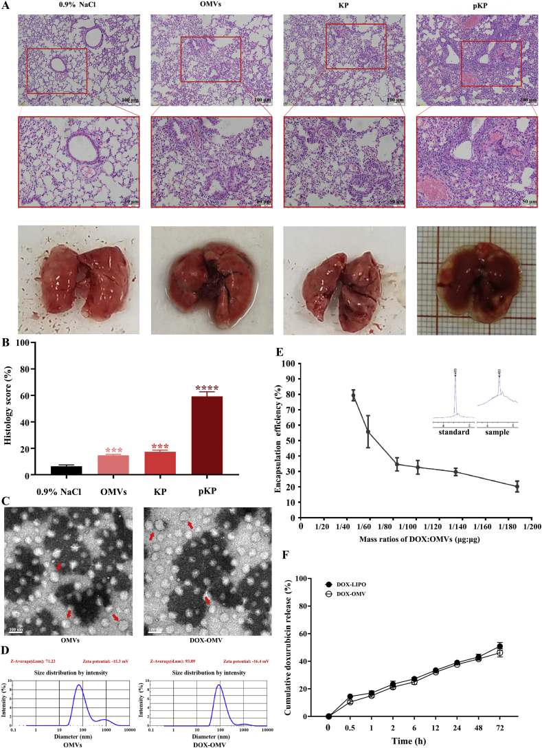
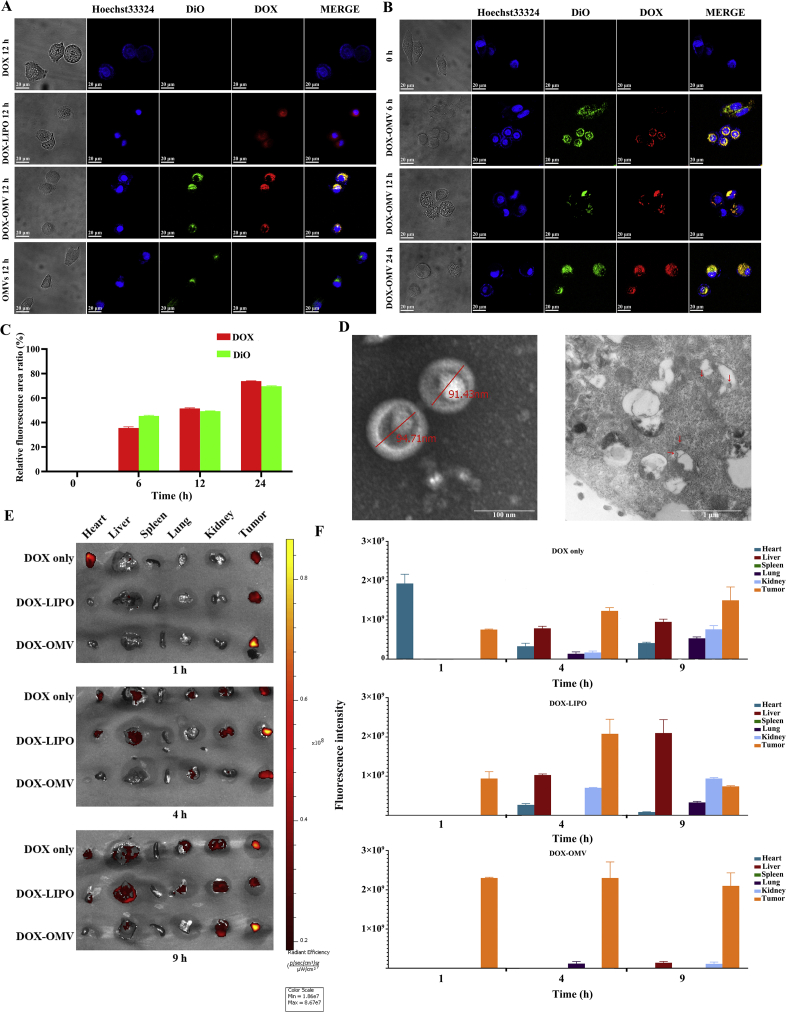
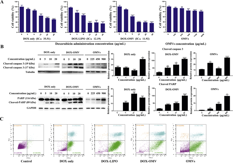
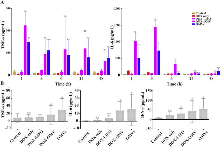
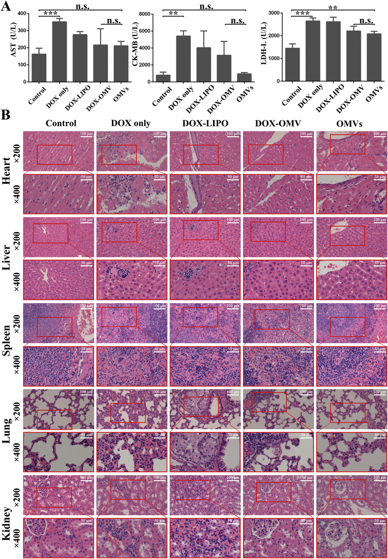
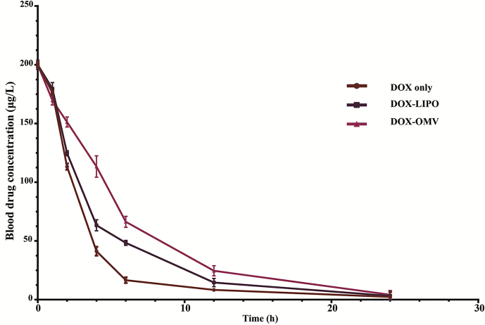
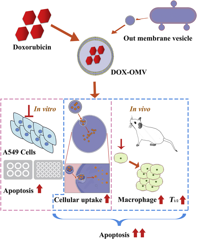
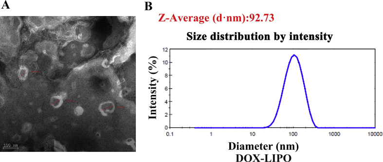
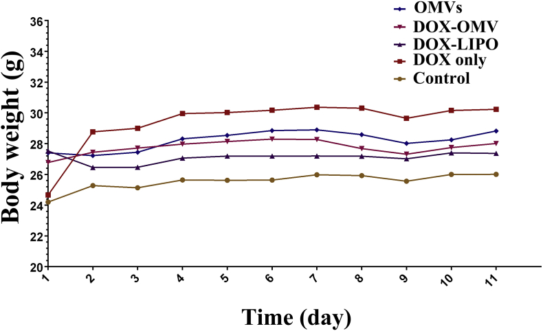
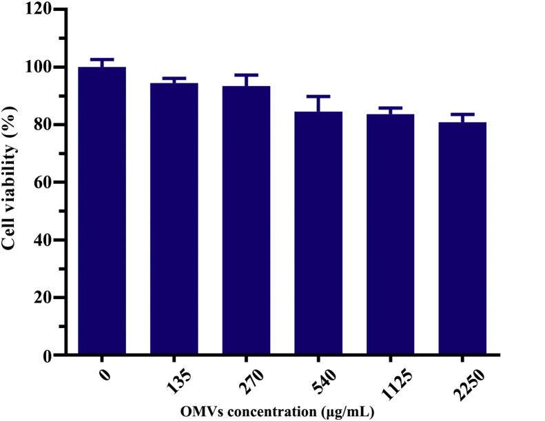
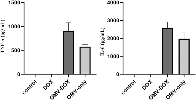
More efficient drug delivery system and formulation with less adverse effects are needed for the clinical application of broad-spectrum antineoplastic agent doxorubicin (DOX). Here we obtained outer-membrane vesicles (OMVs), a nano-sized proteoliposomes naturally released by Gram-negative bacteria, from attenuated Klebsiella pneumonia and prepared doxorubicin-loaded O0MVs (DOX-OMV). Confocal microscopy and in vivo distribution study observed that DOX encapsulated in OMVs was efficiently transported into NSCLC A549 cells. DOX-OMV resulted in intensive cytotoxic effects and cell apoptosis in vitro as evident from MTT assay, Western blotting and flow cytometry due to the rapid cellular uptake of DOX. In A549 tumor-bearing BALB/c nude mice, DOX-OMV presented a substantial tumor growth inhibition with favorable tolerability and pharmacokinetic profile, and TUNEL assay and H&E staining displayed extensive apoptotic cells and necrosis in tumor tissues. More importantly, OMVs' appropriate immunogenicity enabled the recruitment of macrophages in tumor microenvironment which might synergize with their cargo DOX in vivo. Our results suggest that OMVs can not only function as biological nanocarriers for chemotherapeutic agents but also elicit suitable immune responses, thus having a great potential for the tumor chemoimmunotherapy.

Keywords: Anti-tumor efficacy; Bacterial outer-membrane vesicles; Chemoimmunotherapy; Doxorubicin; Non-small-cell lung cancer.


Figures

Image 1
Graphical abstract
Figure 1
Figure 1
Preparation and characterization of DOX-OMV. (A) Virulence of attenuated K. pneumonia ACCC 60095 and its OMVs. BALB/c mice underwent nasal administration of attenuated K. pneumonia (KP) and its OMVs with normal saline as negative control and pathogenic strain (pKP) as positive control once a day for 10 days. Lung tissues were removed for H&E staining. Scale bar = 100 μm (upper), 50 μm (lower). (B) Histology score evaluation of H&E staining by ImageJ software. Data are mean ± SD, n = 3; ∗∗∗P < 0.001, ∗∗∗∗P < 0.0001 vs. negative control. (C) Transmission electron micrographs of OMVs prepared from attenuated K. pneumonia and DOX-loaded OMVs (DOX-OMV). Arrows indicated typical OMVs and DOX-OMV. Scale bar = 100 nm. (D) Size distribution profile of OMVs and DOX-OMV. (E) The encapsulation efficiency (EE) analyzed by LC–MS. Data are mean ± SD, n = 3. (F) Time course of DOX release from DOX-OMV. Data are mean ± SD, n = 3. Drug release study in vitro was carried out in pH7.4 PBS containing 1% (v/v) Tween 80. The DOX-OMV solution was put into the dialysis bag. Next, the dialysis bag was placed in 200 mL of PBS, and then shake incubated at 100 rpm and 37 °C. At different time points, the PBS was sampled for analysis of drug concentration.
Figure 2
Figure 2
Drug uptake by tumor cells. (A)–(C) Cellular uptake of DOX detected by confocal microscopy. A549 cells were treated with (A) OMVs, DOX (20 μg/mL), 20 μg/mL of DOX loaded in OMVs (DOX-OMV) or loaded in liposome (DOX-LIPO) separately for 12 h or (B) DOX-OMV for 0, 6, 12 and 24 h. OMVs were labeled with DiO (green fluorescence). The blue fluorescence represented the location of cell nuclei and the red was the intrinsic fluorescence of DOX. (C) The quantitative analysis of Fig. 2B performed by ImageJ software. The results are calculated as follows: Relative fluorescence area ratio (%) = (The area with green or red fluorescence/The area with blue fluorescence) × 100. Data are mean ± SD, n = 3. (D) TEM images (JEM1230, JEOL, Japan) of DOX-OMV (left) and A549 cells treated with DOX-OMV for 12 h (right). Possible intracellular OMVs were visible in and around vacuoles (Arrows indicate OMVs). (E) and (F) In vivo drug distribution of DOX-OMV in major organs. Mice bearing A549 tumors (∼100 mm3) were injected a single dose of free DOX (2 μL/g), equivalent DOX loaded into OMVs or loaded into liposomes. At 1, 4 and 9 h, mice were selected randomly to obtain tumors and major organs for the investigation of DOX distribution and tumor targeting through ex vivo fluorescence imaging (E). Statistical analysis of fluorescence intensity (data are mean ± SD; n = 3) (F).
Figure 3
Figure 3
In vitro antitumor effect of DOX-OMVs. (A) A549 cells were treated with indicated concentrations of free DOX, equivalent DOX encapsulated in OMVs (DOX-OMVs) or in liposome (DOX-LIPO), and empty OMVs for 24 h. Cell viability was measured by MTT assay (IC50 of DOX, DOX-OMV and DOX-LIPO were 35.51, 12.19 and 11.92 μg/mL, respectively). The data were displayed as mean ± SD, n = 5. (B) After A549 cells were treated with indicated concentrations of OMVs, free DOX or equivalent DOX encapsulated in OMVs (DOX-OMVs), the protein levels of cleaved-caspase 3, PARP and cleaved-PARP were analyzed by Western blotting. Quantitative evaluation of the resulting bands was performed by ImageJ software. (C) A549 cells were treated with 20 μg/mL of free DOX, equivalent DOX encapsulated in OMVs (DOX-OMV) or in liposome (DOX-LIPO), and empty OMVs for 24 h, then cell apoptosis was detected by flow cytometry. ∗P < 0.05, ∗∗P < 0.01, ∗∗∗P < 0.001, ∗∗∗∗P < 0.0001 vs. control, n = 3.
Figure 4
Figure 4
In vivo antitumor efficacy of DOX-OMV. Tumor growth suppression was investigated in A549 xenograft BALB/c mice. Mice bearing tumors (∼50 mm3) were treated (i.p. every day) with PBS, free DOX (2 mg/kg), an equivalent dose of DOX loaded into either OMVs (DOX-OMV) or liposomes (DOX-LIPO), and empty OMVs for 11 days. (A) Mean tumor volume of 5 groups. Data were displayed as mean ± SD, n = 3; ∗∗∗∗P < 0.001 vs. control. (B) The photo of the tumor tissues removed from mice after 11 days' treatment with vehicle control (n = 3), free DOX (n = 3), DOX-OMV (n = 4), DOX-LIPO (n = 4) and empty OMVs (n = 3). Red boxes represent tumors disappeared in mice. (C) TUNEL assay for cell apoptosis (green fluorescence) of tumor tissues. (D) The quantitative analysis of apoptotic cells in TUNEL assay (ImageJ software). Data were displayed as mean ± SD, n = 3; ∗∗∗P < 0.001 vs. control. (E) H&E staining of tumor tissue sections. (F) Immunohistochemical staining for detecting F4/80 glycoprotein in tumor sections. (G) The quantitative evaluation of immunopositive areas (ImageJ software). Data were displayed as mean ± SD, n = 3; ∗∗P < 0.01 vs. control. Scale bar = 20 μm.
Figure 5
Figure 5
Serum immune cytokine analysis in C57BL/6 mice. The mice were treated (i.p. every day) with PBS, free DOX (2 mg/kg), an equivalent dose of DOX loaded into either OMVs (DOX-OMV) or liposomes (DOX-LIPO), and empty OMVs. (A) Blood sample was collected at 1, 3, 6, 24 and 48 h after initial administration, then the levels of TNF-α and IL-6 were analyzed by using respective ELISA kits. (B) After 11 days' repeated treatment, blood samples were collected and detected TNF-α, IL-6 and IFN-γ by respective ELISA kits. Data were displayed as mean ± SD, n = 3. ∗P < 0.05, ∗∗P < 0.01 vs. control. n.s. not significant.
Figure 6
Figure 6
The safety evaluation of DOX-OMV. The tumor bearing BALB/c mice were treated as described in Fig. 4. After 11 days of treatment, various organs including heart, liver, spleen, lung and kidney together with blood samples were collected for further analysis. (A) The levels of serum biomarkers of cardiac damage including AST, CK-MB and LDH. Each bar represented mean ± SD, n = 3; ∗∗P < 0.01, ∗∗∗P < 0.001. n.s. not significant. (B) H&E staining of main organ sections.
Figure 7
Figure 7
Pharmacokinetic profile of DOX-OMVs in rabbits. Animals were administered i.v. at doses of 1.0 mg/kg of free DOX, DOX entrapped in liposomes (DOX-LIPO) or in OMVs (DOX-OMV). Blood samples were taken at 0, 1, 2, 4, 6, 12 and 24 h after drug administration. Data were presented as mean ± SD; n = 3. Pharmacokinetic parameters were calculated by using DAS 2.0 software and shown in Table 1.
Figure 8
Figure 8
Overview of anti-NSCLC effect triggered by DOX-OMV. In vitro, DOX encapsulated in OMVs was efficiently delivered into A549 cells, thus resulted in intensive cytotoxic effects and cell apoptosis. In vivo, OMVs not only functioned as drug delivery carriers but also induced the recruitment of macrophages in tumor microenvironment which might synergize with their cargo DOX.
figs1
figs1
figs2
figs2
figs3
figs3
figs4
figs4
figs5
figs5

References

    1. Bray F., Ferlay J., Soerjomataram I., Siegel R.L., Torre L.A., Jemal A. Global cancer statistics 2018: GLOBOCAN estimates of incidence and mortality worldwide for 36 cancers in 185 countries. Ca - Cancer J Clin. 2018;68:394–424. - PubMed
    1. Zappa C., Mousa S.A. Non-small cell lung cancer: current treatment and future advances. Transl Lung Cancer Res. 2016;5:288–300. - PMC - PubMed
    1. Tacar O., Sriamornsak P., Dass C.R. Doxorubicin: an update on anticancer molecular action, toxicity and novel drug delivery systems. J Pharm Pharmacol. 2013;65:157–170. - PubMed
    1. O’Brien M.E., Wigler N., Inbar M., Rosso R., Grischke E., Santoro A., Catane R.D., Tomczak P., Orlandi F., Mellars L., Alland L., TendlerM C., O'Brien E., Wigler N., Inbar M., Rosso R., Grischke E., Santoro A., Catane R., Kieback D.G., Tomczak P., Ackland S.P., Orlandi F., Mellars L., Alland L., Tendler C. Reduced cardiotoxicity and comparable efficacy in a phase III trial of pegylated liposomal doxorubicin HCl (CAELYX™/Doxil®) versus conventional doxorubicin for first-line treatment of metastatic breast cancer. Ann Oncol. 2004;15:440–449. - PubMed
    1. Barenholz Y. Doxil® — the first FDA-approved nano-drug: lessons learned. J Contr Release. 2012;160:117–134. - PubMed

LinkOut - more resources