Review
. 2020 Sep 14;6(3):00289-2020.
doi: 10.1183/23120541.00289-2020. eCollection 2020 Jul.

Pulmonary alveolar microlithiasis: no longer in the stone age

Affiliations
Review

Pulmonary alveolar microlithiasis: no longer in the stone age

Elisabeth Bendstrup et al. ERJ Open Res. .

Abstract

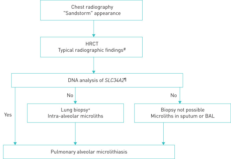
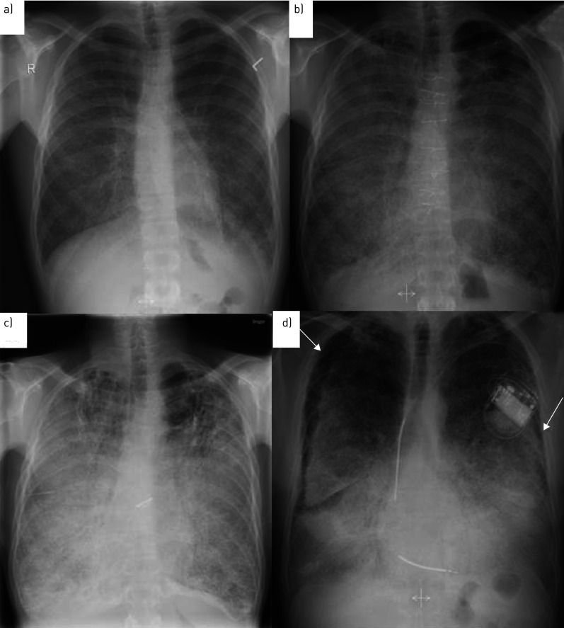
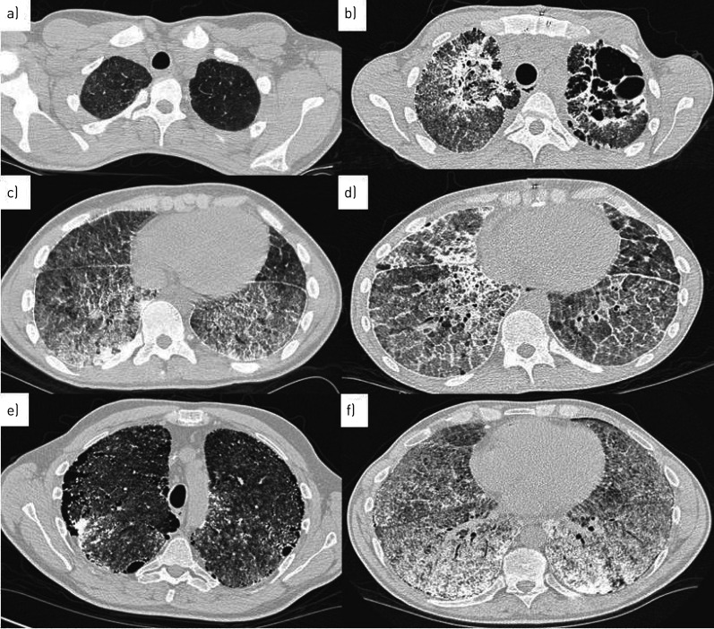
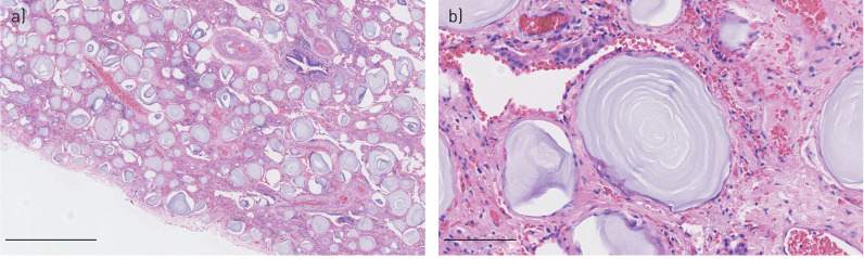
Pulmonary alveolar microlithiasis (PAM) is a rare parenchymal lung disease caused by variants in the SCL34A2 gene and characterised by the accumulation of intra-alveolar microliths. PAM has been reported in fewer than 1100 cases throughout the world. It is an autosomal recessive hereditary disease and often associated with consanguinity. Progress with respect to the genetic background and pathophysiology has resulted in an increased understanding of the disease in recent years. Until now, 30 genetic different SLC34A2 variants have been reported, which all are considered significant for disease development. There is no sex difference and the majority of cases are diagnosed at the age of 30-40 years. Many patients are asymptomatic and the diagnosis is made at random. When symptomatic, dyspnoea, cough, chest pain and fatigue are common complaints. The diagnosis of PAM can confidently be based on typical radiographic findings and genetic testing proving rare biallelic SCL34A2 gene variants. Bronchoalveolar lavage and histopathology may show microliths. There is no disease-specific treatment and management is supportive. Lung transplantation should be considered in advanced cases.


Conflict of interest statement

Conflict of interest: E. Bendstrup has nothing to disclose. Conflict of interest: Å.L.M. Jönsson has nothing to disclose.

Figures

FIGURE 1
FIGURE 1
Diagnostic algorithm for pulmonary alveolar microlithiasis. The algorithm is step-wise with the least invasive procedure preferred as the first choice. The decision to obtain tissue should always be individualised and preferably be taken at a multidisciplinary team discussion. Less experienced centres are recommended to consult an expert centre. #: intra-alveolar calcifications, micronodules, ground-glass opacities, thickened septa, subpleural cysts; : if genetic analysis is not available, proceed to invasive examinations;+: transbronchial forceps or cryobiopsy; the choice between these should be based on availability and experience and considering the least invasive procedure first; if transbronchial biopsies are non-diagnostic, consider surgical lung biopsy.
FIGURE 2
FIGURE 2
Chest radiography of two patients with pulmonary alveolar microlithiasis. a) Patient 1, 2003, radiological severity stage II. b) Patient 1, 2013, radiological severity stage III. c) Patient 1, 2019, radiological severity stage IV. d) Patient 2, 2007, radiological severity stage IV. White arrows demonstrate the “black pleural sign”.
FIGURE 3
FIGURE 3
High-resolution computed tomography of two patients with pulmonary alveolar microlithiasis. a) Patient 1, 2003, ground-glass opacities in apical lung fields and c) calcifications, micronodules, thickened septa in basal lung fields. b) Patient 1, 2016, calcifications with micronodules, thickened septa and cysts in apical lung fields and d) progression of calcifications with consolidations and crazy paving. e) Patient 2, 2005, radiological severity stage IV. Subpleural microcysts (black pleural sign), ground-glass opacities and micronodules in apical lung fields and f) same patient, basal lung fields showing extensive calcifications with thickened septa and micronodules.
FIGURE 4
FIGURE 4
Lung section from a male patient with pulmonary alveolar microlithiasis showing calcified microliths in the intra-alveolar spaces. Haematoxylin and eosin staining. Original magnification: a) ×50 and b) ×400. Scale bars: a) 1 mm and b) 100 μm. By courtesy of pathologist Johanne Lade Keller, Dept of Pathology, Aarhus University Hospital, Aarhus, Denmark.

References

    1. Francisco FA, Pereira e Silva JL, Hochhegger B, et al. . Pulmonary alveolar microlithiasis. State-of-the-art review. Respir Med 2013; 107: 1–9. doi:10.1016/j.rmed.2012.10.014 - DOI - PubMed
    1. Anzalone M, ed Marcello Malpighi e i suoi scritti sugli organi del respire. Manoscr vol XII, foglio 81 Bologna, Tamari Editori, 1966: pp. 259–262.
    1. Harbitz F. Extensive calcification of the lungs as a distinct disease. Arch Intern Med 1918; 21: 139–146. doi:10.1001/archinte.1918.00090070150012 - DOI
    1. Puhr I. Microlithiasis alveolaris pulmonum. Virchows Arch A Patho Anat Histopathol 1933; 290: 156. doi:10.1007/BF01979722 - DOI
    1. Khaladkar SM, Kondapavuluri SK, Kamal A, et al. . Pulmonary alveolar microlithiasis - clinico-radiological dissociation - A case report with radiological review. J Radiol Case Rep 2016; 10: 14–21. - PMC - PubMed

LinkOut - more resources