. 2020 Sep 14;6(3):00210-2019.
doi: 10.1183/23120541.00210-2019. eCollection 2020 Jul.

Airway-artery quantitative assessment on chest computed tomography in paediatric primary ciliary dyskinesia

Affiliations

Airway-artery quantitative assessment on chest computed tomography in paediatric primary ciliary dyskinesia

Valentina Ferraro et al. ERJ Open Res. .

Abstract

Chest computed tomography (CT) is the gold standard for detecting structural abnormalities in patients with primary ciliary dyskinesia (PCD) such as bronchiectasis, bronchial wall thickening and mucus plugging. There are no studies on quantitative assessment of airway and artery abnormalities in children with PCD. The objectives of the present study were to quantify airway and artery dimensions on chest CT in a cohort of children with PCD and compare these with control children to analyse the influence of covariates on airway and artery dimensions. Chest CTs of 13 children with PCD (14 CT scans) and 12 control children were collected retrospectively. The bronchial tree was segmented semi-automatically and reconstructed in a three-dimensional view. All visible airway-artery (AA) pairs were measured perpendicular to the airway centre line, annotating per branch inner and outer airway and adjacent artery diameter and computing inner airway diameter/artery ratio (AinA ratio), outer airway diameter/artery ratio (AoutA ratio), wall thickness (WT), WT/outer airway diameter ratio (Awt ratio) and WT/artery ratio. In the children with PCD (38.5% male, mean age 13.5 years, range 9.8-15.3) 1526 AA pairs were measured versus 1516 in controls (58.3% male, mean age 13.5 years, range 8-14.8). AinA ratio and AoutA ratio were significantly higher in children with PCD than in control children (both p<0.001). Awt ratio was significantly higher in control children than in children with PCD (p<0.001). Our study showed that in children with PCD airways are more dilated than in controls and do not show airway wall thickening.


Conflict of interest statement

Conflict of interest: V. Ferraro has nothing to disclose. Conflict of interest: E-R. Andrinopoulou has nothing to disclose. Conflict of interest: A.M.M. Sijbring has nothing to disclose. Conflict of interest: E.G. Haarman has nothing to disclose. Conflict of interest: H.A.W.M. Tiddens reports unconditional research grants outside the submitted work from Roche, Novartis, CFF, Vertex, Chiesi, Vectura and Gilead; he has participated in the last 5 years in expert panels for Vertex and Gilead. In addition, Dr Tiddens has a patent licensed for the PRAGMA-CF scoring system. He heads the Erasmus MC–Sophia Children's Hospital core laboratory Lung Analysis. FLUIDDA has developed computational fluid dynamic modelling based on chest CTs obtained from Erasmus MC–Sophia for which royalties are received by Sophia Research BV. All financial aspects for the grants are handled by Sophia Research BV. Conflict of interest: M.W.H. Pijnenburg has nothing to disclose.

Figures

FIGURE 1
FIGURE 1
a) Screen shot of airway tree three-dimensional reconstruction. b) Three measurements of the edge of airway and accompanying artery were used to annotate surface areas of the inner (A) and outer (B) airway and of the artery (C) in order to estimate diameters and to calculate the airway–artery ratios.
FIGURE 2
FIGURE 2
Anatomy of the airways. a) Airway generations are defined with the numbers in different coloured segmental branches; each segmental branch starts with a different generation number (i.e. 3–4 in the upper segmental branches and 5–7 in the lower segmental branches). b) In order to correct the discrepancy between upper and lower segmental branches, we renumbered each segmental generation by labelling the first segmental bronchi as generation 1 (Artist: K. Rubenis) [21, 22].
FIGURE 3
FIGURE 3
Flowchart of primary ciliary dyskinesia (PCD) patients included. CT: computed tomography.
FIGURE 4
FIGURE 4
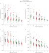
Box plots of a) inner airway diameter, b) outer airway diameter, c) wall thickness and d) artery diameter of primary ciliary dyskinesia (PCD) and control patients, divided according to different segmental generation. Each box shows median (horizontal line), interquartile range (solid box), 1.5×interquartile range (whiskers) and outliers (points and stars). *: statistically significant (p<0.05).
FIGURE 5
FIGURE 5
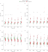
Box plots of airway–artery ratio of primary ciliary dyskinesia (PCD) and control patients, divided according to different segmental generation. Each box shows median (horizontal line), interquartile range (solid box), 1.5×interquartile range (whiskers) and outliers (points and stars). AinA: inner airway diameter/artery diameter; AoutA: outer airway diameter/artery diameter; Awt: airway wall thickness/outer airway diameter; AwtA: airway wall thickness/artery diameter. *: statistically significant (p<0.05).
FIGURE 6
FIGURE 6
Box plots of airway–artery ratio of primary ciliary dyskinesia (PCD) and control patients, divided according to different lung lobes. Each box shows median (horizontal line), interquartile range (solid box), 1.5×interquartile range (whiskers) and outliers (points and stars). AinA: inner airway diameter/artery diameter; AoutA: outer airway diameter/artery diameter; Awt: airway wall thickness/outer airway diameter; AwtA: airway wall thickness/artery diameter. *: statistically significant (p<0.05).

References

    1. Lucas JS, Alanin MC, Collins S, et al. . Clinical care of children with primary ciliary dyskinesia. Expert Rev Respir Med 2017; 11: 779–790. doi:10.1080/17476348.2017.1360770 - DOI - PubMed
    1. Lucas JS, Chetcuti P, Copeland F, et al. . Overcoming challenges in the management of primary ciliary dyskinesia: the UK model. Paediatr Respir Rev 2014; 15: 142–145. - PubMed
    1. Shapiro AJ, Zariwala MA, Ferkol T, et al. . Diagnosis, monitoring, and treatment of primary ciliary dyskinesia: PCD foundation consensus recommendations based on state-of-the-art review. Pediatr Pulmonol 2016; 51: 115–132. doi:10.1002/ppul.23304 - DOI - PMC - PubMed
    1. Goutaki M, Meier AB, Halbeisen FS, et al. . Clinical manifestations in primary ciliary dyskinesia: systematic review and meta-analysis. Eur Respir J 2016; 48: 1081–1095. doi:10.1183/13993003.00736-2016 - DOI - PubMed
    1. Mullowney T, Manson D, Kim R, et al. . Primary ciliary dyskinesia and neonatal respiratory distress. Pediatrics 2014; 134: 1160–1166. doi:10.1542/peds.2014-0808 - DOI - PMC - PubMed

LinkOut - more resources