

Variable echo time imaging for detecting the short T2* components of the sciatic nerve: a validation study
- PMID: 32964300
- PMCID: PMC8154754
- DOI: 10.1007/s10334-020-00886-w
Variable echo time imaging for detecting the short T2* components of the sciatic nerve: a validation study
Abstract
Objective: The aim of this study was to develop and validate an MRI protocol based on a variable echo time (vTE) sensitive to the short T2* components of the sciatic nerve.
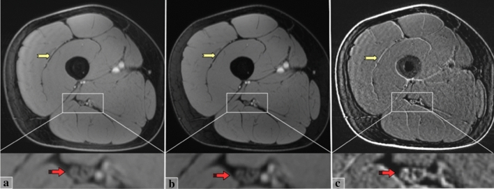
Materials and methods: 15 healthy subjects (M/F: 9/6; age: 21-62) were scanned at 3T targeting the sciatic nerve at the thigh bilaterally, using a dual echo variable echo time (vTE) sequence (based on a spoiled gradient echo acquisition) with echo times of 0.98/5.37 ms. Apparent T2* (aT2*) values of the sciatic nerves were calculated with a mono-exponential fit and used for data comparison.
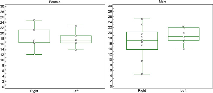
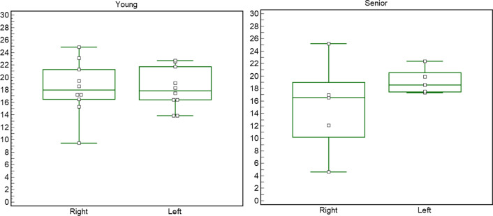
Results: There were no significant differences in aT2* related to side, sex, age, and BMI, even though small differences for side were reported. Good-to-excellent repeatability and reproducibility were found for geometry of ROIs (Dice indices: intra-rater 0.68-0.7; inter-rater 0.70-0.72) and the related aT2* measures (intra-inter reader ICC 0.95-0.97; 0.66-0.85) from two different operators. Side-related signal-to-noise-ratio non-significant differences were reported, while contrast-to-noise-ratio measures were excellent both for side and echo.
Discussion: Our study introduces a novel MR sequence sensitive to the short T2* components of the sciatic nerve and may be used for the study of peripheral nerve disorders.
Keywords: Fibrosis; Magnetic resonance imaging; Peripheral nerves; Validation study.
Conflict of interest statement
The authors declare that they have no conflict of interest.
Figures
Similar articles
-
T2 mapping of the distal sciatic nerve in healthy subjects and patients suffering from lumbar disc herniation with nerve compression.MAGMA. 2020 Oct;33(5):713-724. doi: 10.1007/s10334-020-00832-w. Epub 2020 Feb 11. MAGMA. 2020. PMID: 32048099 Free PMC article.
-
Feasibility of dynamic T2 *-based oxygen-enhanced lung MRI at 3T.Magn Reson Med. 2024 Mar;91(3):972-986. doi: 10.1002/mrm.29914. Epub 2023 Nov 27. Magn Reson Med. 2024. PMID: 38013206 Free PMC article.
-
Magnetic resonance imaging evaluation of acute crush injury of rabbit sciatic nerve: correlation with histology.Can Assoc Radiol J. 2008 Jun;59(3):123-30. Can Assoc Radiol J. 2008. PMID: 18697718
-
Dual-Echo Turbo Spin Echo and 12-Echo Multi Spin Echo Sequences as Equivalent Techniques for Obtaining T2-Relaxometry Data: Application in Symptomatic and Asymptomatic Hereditary Transthyretin Amyloidosis as a Surrogate Disease.Invest Radiol. 2022 May 1;57(5):301-307. doi: 10.1097/RLI.0000000000000837. Invest Radiol. 2022. PMID: 34839307
-
Ultrashort echo time (UTE) magnetic resonance imaging of the short T2 components in white matter of the brain using a clinical 3T scanner.Neuroimage. 2014 Feb 15;87:32-41. doi: 10.1016/j.neuroimage.2013.10.053. Epub 2013 Nov 2. Neuroimage. 2014. PMID: 24188809 Free PMC article.
Cited by
-
Quantitative MRI Assessment Using Variable Echo Time Imaging of Peripheral Nerve Injury in ATTRv Amyloidosis Patients.Eur J Neurol. 2025 Apr;32(4):e70172. doi: 10.1111/ene.70172. Eur J Neurol. 2025. PMID: 40265689 Free PMC article.
References
-
- Pichiecchio A, Rossi M, Cinnante C, Colafati GS, De Icco R, Parini R, Menni F, Furlan F, Burlina A, Sacchini M, Donati MA, Fecarotta S, Casa RD, Deodato F, Taurisano R, Di Rocco M. Muscle MRI of classic infantile pompe patients: Fatty substitution and edema-like changes. Muscle Nerve. 2017 doi: 10.1002/mus.25417. - DOI - PubMed
-
- Chhabra A, Andreisek G. Magnetic resonance neurography. New Delhi: Jaypee Brothers Medical Pub; 2012.
MeSH terms
Grants and funding
LinkOut - more resources
Full Text Sources
Medical
