. 2020 Nov;98(11):1639-1656.
doi: 10.1007/s00109-020-01974-z. Epub 2020 Sep 22.

Fibulin-3 knockout mice demonstrate corneal dysfunction but maintain normal retinal integrity

Affiliations

Fibulin-3 knockout mice demonstrate corneal dysfunction but maintain normal retinal integrity

Steffi Daniel et al. J Mol Med (Berl). 2020 Nov.

Abstract

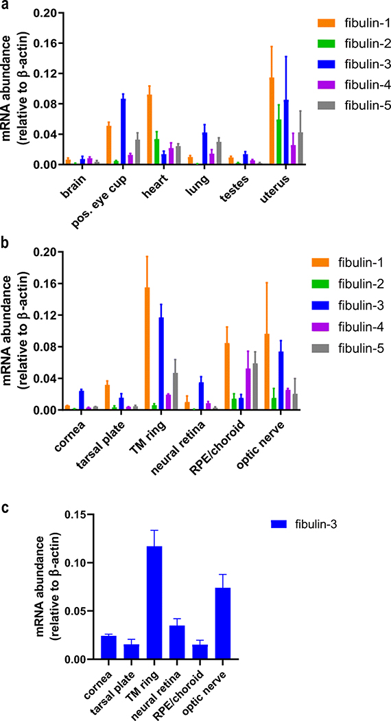
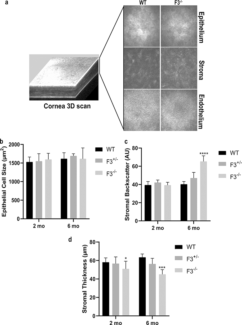
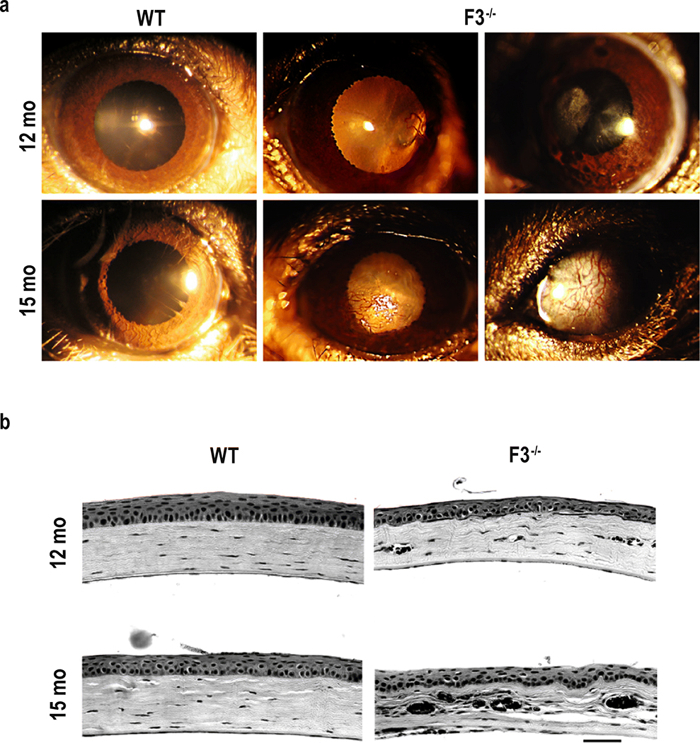
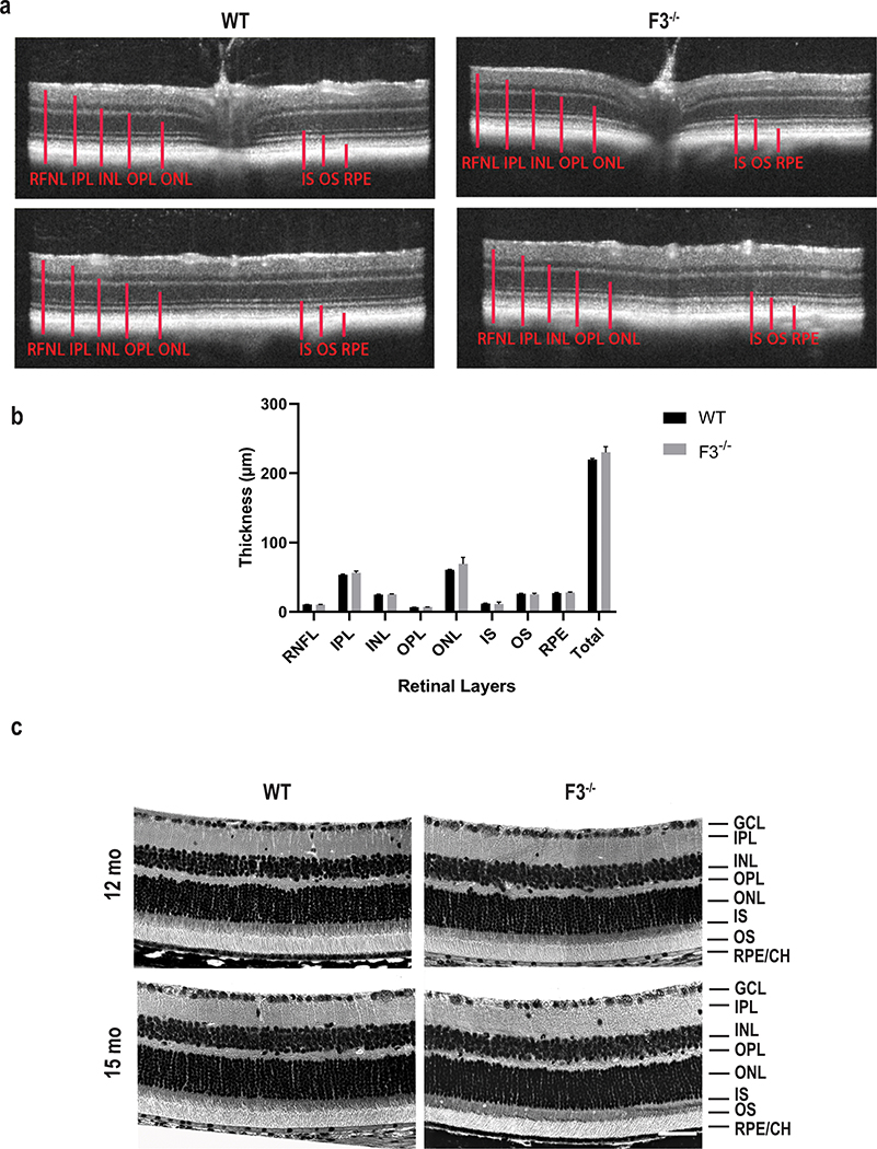
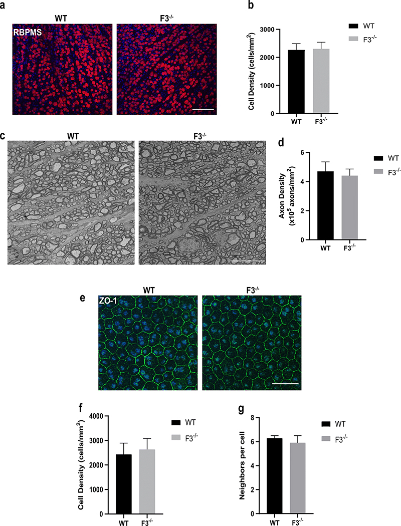
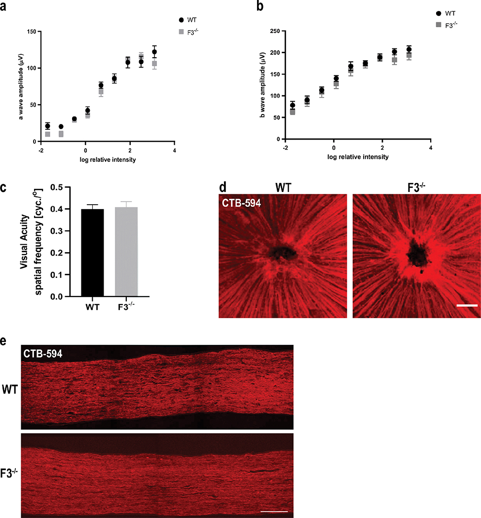
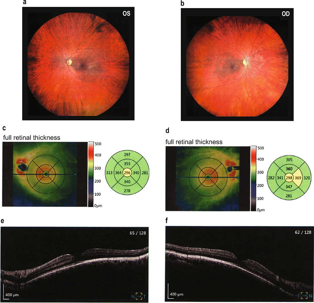
Fibulin-3 (F3) is an extracellular matrix glycoprotein found in basement membranes across the body. An autosomal dominant R345W mutation in F3 causes a macular dystrophy resembling dry age-related macular degeneration (AMD), whereas genetic removal of wild-type (WT) F3 protects mice from sub-retinal pigment epithelium (RPE) deposit formation. These observations suggest that F3 is a protein which can regulate pathogenic sub-RPE deposit formation in the eye. Yet the precise role of WT F3 within the eye is still largely unknown. We found that F3 is expressed throughout the mouse eye (cornea, trabecular meshwork (TM) ring, neural retina, RPE/choroid, and optic nerve). We next performed a thorough structural and functional characterization of each of these tissues in WT and homozygous (F3-/-) knockout mice. The corneal stroma in F3-/- mice progressively thins beginning at 2 months, and the development of corneal opacity and vascularization starts at 9 months, which worsens with age. However, in all other tissues (TM, neural retina, RPE, and optic nerve), gross structural anatomy and functionality were similar across WT and F3-/- mice when evaluated using SD-OCT, histological analyses, electron microscopy, scotopic electroretinogram, optokinetic response, and axonal anterograde transport. The lack of noticeable retinal abnormalities in F3-/- mice was confirmed in a human patient with biallelic loss-of-function mutations in F3. These data suggest that (i) F3 is important for maintaining the structural integrity of the cornea, (ii) absence of F3 does not affect the structure or function of any other ocular tissue in which it is expressed, and (iii) targeted silencing of F3 in the retina and/or RPE will likely be well-tolerated, serving as a safe therapeutic strategy for reducing sub-RPE deposit formation in disease. KEY MESSAGES: • Fibulins are expressed throughout the body at varying levels. • Fibulin-3 has a tissue-specific pattern of expression within the eye. • Lack of fibulin-3 leads to structural deformities in the cornea. • The retina and RPE remain structurally and functionally healthy in the absence of fibulin-3 in both mice and humans.

Keywords: Age-related macular degeneration (AMD); Cornea; EFEMP1; Fibulin-3; Malattia Leventinese (ML); Retina.


Conflict of interest statement

Conflict of interest

The authors declare that they have no conflicts of interest.

Figures

Fig. 1
Fig. 1
Gene expression profile of the fibulins. (a) Expression levels of fibulin 1–5 in brain, posterior (pos.) eye cup, heart, lung, testes, and uterus relative to β-actin. (b) Expression levels of fibulin 1–5 in ocular tissues, namely cornea, tarsal plate, trabecular meshwork (TM) ring, neural retina, RPE/choroid, and optic nerve relative to β-actin. c Reiteration of Fig. 1b focusing on the expression of F3 in ocular tissues. The values are represented as mean ± SD (n = 3–5)
Fig. 2
Fig. 2
Lack of F3 leads to stromal thinning as well as increased corneal haze in F3−/− animals. (a) Representative image of in vivo 3D corneal scan. Panels showing comparative images of epithelial, stromal, and endothelial layers of WT and F3−/− animals. (b) Graph representing epithelial cell size (μm2) shows no differences between WT, F3+/−, and F3−/− in 2 month (n = 7–12) and 6 month (n = 4–7) animals. (c) Graph representing stromal backscatter (AU) displays no differences between WT, F3+/−, and F3−/− at 2 months (n = 7–12), while increased stromal backscatter is observed in F3−/− animals compared with that in WT at 6 months (n = 4–7, p < 0.0001). (d) Graph representing stromal thickness (μm) shows decreased thickness in F3−/− at 2months (n = 7–12, p = 0.02) and 6 months (n = 4–7, p < 0.001) compared with that in WT. Analyses performed by 2-way ANOVA and represented as mean ± SD
Fig. 3
Fig. 3
Corneal opacification, vascularization, and deterioration in F3−/− mice. (a) Representative images of slit-lamp examination. Panels depict various phenotypic manifestations of corneal changes in 12 months and 15 months F3−/− animals compared with WT (n = 4–6). (b) Corneal histology. Representative images showing histological changes in the cornea of F3−/− animals compared with that of WT at 12 months and 15 months (n = 4–5) (scale bar = 50 μm)
Fig. 4
Fig. 4
Retina remains structurally intact in the absence of F3. (a) Representative images of SD-OCT examination. SD-OCT scans of F3−/− and WT mice show no distinguishable changes in thickness for all the retinal layers at 9 months. Top panel represents scans from optic nerve head and bottom panels from the periphery. (b) Graph representing comparison of thickness of retinal layers between WT and F3−/− represented as mean ± SD. (c) Retinal histology. Representative images showing comparable histological structures in F3−/− and WT animals at 15 months (scale bar = 50 μm). RNFL retinal nerve fiber layer, GCL ganglion cell layer, IPL inner plexiform layer, INL inner nuclear layer, OPL outer plexiform layer, ONL outer nuclear layer, IS inner segment, OS outer segment, RPE retinal pigment epithelium, CH choroid (n = 6–7)
Fig. 5
Fig. 5
Ganglion cells, axons, and RPE appear unaffected in F3−/− animals. (a) Representative images of RGCs stained with RBPMS in WT and F3−/− mice at 15 months (scale bar = 100 μm). (b) Graph representing cell density (cells/mm2) shows no differences between WT and F3−/− 15 month mice (n = 5–6). (c) Representative images of optic nerve cross sections after electron microscopy of WT and F3−/− mice at 15 months (scale bar = 10 μm). (d) Graphical representation of axonal density (×105 axons/mm2) shows no difference between WT and F3−/− (n = 3–4). (e) ZO-1 staining of RPE cells of WT and F3−/− in 15 month mice (scale bar = 100 μm). (f) RPE cell density (cells/mm2). (g) Number of neighboring cells are similar in comparison for WT and F3−/− animals (n = 5–6). Analyses performed by 2-way ANOVA and represented as mean ± SD
Fig. 6
Fig. 6
Normal retinal physiology and function are observed in the absence of F3. (a) An ERG exam revealed no significant differences in a-wave response (b) or b-wave response between WT and F3−/− animals at 6 months (n = 10–12 eyes). (c) Normal visual acuity was recorded for both WT and F3−/− animals by OKR at 2 months (n = 5). Analyses performed by 2-way ANOVA and represented as mean ± SD. (d) Representative images of CTB-594 tracer through nerve fiber layer (scale bar = 100 μm) and (e) optic nerve axons did not show any transport deficit in F3−/− mice compared with WT at 15 months (scale bar = 100 μm)
Fig. 7
Fig. 7
Normal retinal structure in a patient with biallelic loss-of-function mutations in F3. (a, b) Unremarkable fundus photographs from a patient that has mutations (p.(Met107fs, resulting in a premature stop codon at residue 127) and p.(Tyr205*)) on separate alleles encoding for F3. (c–f) OCT-mediated measurements demonstrate normal retinal thickness (average of 305 μm OS, 310 μm OD) and total retinal volume (8.64 mm3 OS, 8.77 mm3 OD)

Similar articles

Cited by

References

    1. Zhang Y, Marmorstein LY (2010) Focus on molecules: fibulin-3 (EFEMP1). Exp Eye Res 90:374–375 - PMC - PubMed
    1. Hulleman JD (2016) Malattia Leventinese/Doyne honeycomb retinal dystrophy: similarities to age-related macular degeneration and potential therapies. Adv Exp Med Biol 854:153–158 - PubMed
    1. McLaughlin PJ, Bakall B, Choi J, Liu Z, Sasaki T, Davis EC, Marmorstein AD, Marmorstein LY (2007) Lack of fibulin-3 causes early aging and herniation, but not macular degeneration in mice. Hum Mol Genet 16:3059–3070 - PubMed
    1. Papke CL, Yanagisawa H (2014) Fibulin-4 and fibulin-5 in elastogenesis and beyond: Insights from mouse and human studies. Matrix Biol 37:142–149 - PMC - PubMed
    1. Kumra H, Nelea V, Hakami H, Pagliuzza A, Djokic J, Xu J, Yanagisawa H, Reinhardt DP (2019) Fibulin-4 exerts a dual role in LTBP-4L-mediated matrix assembly and function. Proc Natl Acad Sci U S A 116:20428–20437 - PMC - PubMed

Publication types

LinkOut - more resources