

CT imaging of pulmonary embolism in patients with COVID-19 pneumonia: a retrospective analysis
- PMID: 32964337
- PMCID: PMC7508235
- DOI: 10.1007/s00330-020-07300-y
CT imaging of pulmonary embolism in patients with COVID-19 pneumonia: a retrospective analysis
Abstract
Objectives: To describe imaging and laboratory findings of confirmed PE diagnosed in COVID-19 patients and to evaluate the characteristics of COVID-19 patients with clinical PE suspicion. Characteristics of patients with COVID-19 and PE suspicion who required admission to the intensive care unit (ICU) were also analysed.
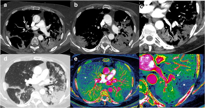
Methods: A retrospective study from March 18, 2020, until April 11, 2020. Inclusion criteria were patients with suspected PE and positive real-time reverse-transcription polymerase chain reaction (RT-PCR) for SARS-CoV-2. Exclusion criteria were negative or inconclusive RT-PCR and other chest CT indications. CTPA features were evaluated and severity scores, presence, and localisation of PE were reported. D-dimer and IL-6 determinations, ICU admission, and previous antithrombotic treatment were registered.
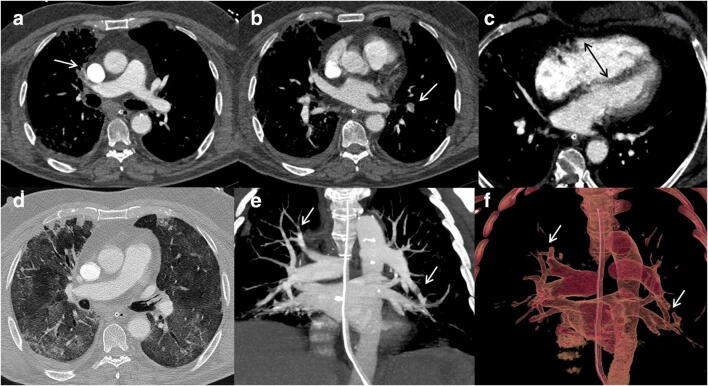
Results: Forty-seven PE suspicions with confirmed COVID-19 underwent CTPA. Sixteen patients were diagnosed with PE with a predominant segmental distribution. Statistically significant differences were found in the highest D-dimer determination in patients with PE and ICU admission regarding elevated IL-6 values.
Conclusion: PE in COVID-19 patients in our series might predominantly affect segmental arteries and the right lung. Results suggest that the higher the D-dimer concentration, the greater the likelihood of PE. Both assumptions should be assessed in future studies with a larger sample size.
Key points: • On CT pulmonary angiography, pulmonary embolism in COVID-19 patients seems to be predominantly distributed in segmental arteries of the right lung, an assumption that needs to be approached in future research. • Only the highest intraindividual determination of d-dimer from admission to CT scan seems to differentiate patients with pulmonary embolism from patients with a negative CTPA. However, interindividual variability calls for future studies to establish cut-off values in COVID-19 patients. • Further studies with larger sample sizes are needed to determine whether the presence of PE could increase the risk of intensive care unit (ICU) admission in COVID-19 patients.
Keywords: COVID-19; Computed tomography angiography; Fibrin fragment D; Intensive care units; Pulmonary embolism.
Conflict of interest statement
The authors of this manuscript declare no relationships with any companies whose products or services may be related to the subject matter of the article.
Figures
References
-
- Bosson JL, Barro C, Satger B, Carpentier PH, Polack B, Pernod G. Quantitative high D-dimer value is predictive of pulmonary embolism occurrence independently of clinical score in a well-defined low risk factor population. J Thromb Haemost. 2005;3:93–99. doi: 10.1111/j.1538-7836.2004.01045.x. - DOI - PubMed
MeSH terms
LinkOut - more resources
Full Text Sources
Medical
Miscellaneous
