Comparative Study
. 2021 Feb;238(2):219-248.
doi: 10.1111/joa.13311. Epub 2020 Sep 22.

Comparative osteology of the hynobiid complex Liua-Protohynobius-Pseudohynobius (Amphibia, Urodela): Ⅰ. Cranial anatomy of Pseudohynobius

Affiliations
Comparative Study

Comparative osteology of the hynobiid complex Liua-Protohynobius-Pseudohynobius (Amphibia, Urodela): Ⅰ. Cranial anatomy of Pseudohynobius

Jia Jia et al. J Anat. 2021 Feb.

Abstract

Hynobiidae are a clade of salamanders that diverged early within the crown radiation and that retain a considerable number of features plesiomorphic for the group. Their evolutionary history is informed by a fossil record that extends to the Middle Jurassic Bathonian time. Our understanding of the evolution within the total group of Hynobiidae has benefited considerably from recent discoveries of stem hynobiids but is constrained by inadequate anatomical knowledge of some extant forms. Pseudohynobius is a derived hynobiid clade consisting of five to seven extant species living endemic to southwestern China. Although this clade has been recognized for over 37 years, osteological details of these extant hynobiids remain elusive, which undoubtedly has contributed to taxonomic controversies over the hynobiid complex Liua-Protohynobius-Pseudohynobius. Here we provide a bone-by-bone study of the cranium in the five extant species of Pseudohynobius (Ps. flavomaculatus, Ps. guizhouensis, Ps. jinfo, Ps. kuankuoshuiensis and Ps. shuichengensis) based on x-ray computer tomography data for 18 specimens. Our results indicate that the cranium in each of these species has a combination of differences in morphology, proportions and articulation patterns in both dermal and endochondral bones. Our study establishes a range of intraspecific differences that will serve as organizing hypotheses for future studies as more extensive collections of these species become available. Morphological features in the cranium for terrestrial ecological adaptation in Hynobiidae are summarized. Based on the results, we also discuss the evolution and development of several potential synapomorphies of Hynobiidae, including features of the orbitosphenoid and articular.

Keywords: Pseudohynobius; Hynobiidae; cranial osteology; development; evolution; salamander; terrestrial adaptation.


Conflict of interest statement

The authors declare there are no competing interests.

Figures

FIGURE 1
FIGURE 1
Interrelationship of species of Pseudohynobius and the LiuaProtohynobiusPseudohynobius complex based on Peng et al. (2010) and Zhao et al. (2016). Note that photographs of representative formaldehyde‐preserved specimens of each species are in dorsal view: Ps. flavomaculatus (the paratype CIB 17343); Ps. jinfo (CIB 85290, the holotype); Ps. guizhouensis (LTHC 0711005, the holotype); Ps. kuankuoshuiensis (ZMC 7604025); Ps. shuichengensis (the paratype LTHC 9460085); Pr. puxiongensis (CIB 98264, the holotype); Liua tsinpaensis (CIB 18349, the holotype); Liua shihi (CIB 19910401); Batrachuperus yenyuanensis (CIB 17005). Ps, Pseudohynobius; Pr, Protohynobius
FIGURE 2
FIGURE 2
Micro‐CT rendered reconstruction of the skull of the five species of Pseudohynobius in dorsal view: (a) Ps. flavomaculatus (CIB 17342, the holotype), (b) Ps. jinfo (CIB 85290, the holotype), (c) Ps. guizhouensis (LTHC 0711005, the holotype), (d) Ps. kuankuoshuiensis (ZMC 7504023, the holotype), (e) Ps. shuichengensis (LTHC 9460084, a paratype). adf, anterodorsal fenestra; app, ascending process of palatoquadrate; fpf, frontoparietal fontanelle; fr, frontal; lac, lacrimal; mx, maxilla; na, nasal; op‐ex, opisthotic‐exoccipital complex; pa, parietal; pm, premaxilla; prf, prefrontal; pro, prootic; pt, pterygoid; qu, quadrate; sm, septomaxilla; sq, squamosal; st, stapes
FIGURE 3
FIGURE 3
Micro‐CT rendered reconstruction of the skull of the five species of Pseudohynobius in ventral view: (a) Ps. flavomaculatus (CIB 17342, the holotype), (b) Ps. jinfo (CIB 85290, the holotype), (c) Ps. guizhouensis (LTHC 0711005, the holotype), (d) Ps. kuankuoshuiensis (ZMC 7504023, the holotype), (e) Ps. shuichengensis (LTHC 9460084, a paratype). amf, anteromedial fenestra; app, ascending process of palatoquadrate; mx, maxilla; obs, orbitosphenoid; op‐ex, opisthotic‐exoccipital complex; pm, premaxilla; pro‐op‐ex, prootic‐opisthotic‐exoccipital complex; ps, parasphenoid; pt, pterygoid; qu, quadrate; st, stapes; vo, vomer
FIGURE 4
FIGURE 4
Micro‐CT rendered reconstruction of the skull of the five species of Pseudohynobius in left lateral view: (a) Ps. flavomaculatus (CIB 17342, the holotype), (b) Ps. jinfo (CIB 85290, the holotype), (c) Ps. guizhouensis (LTHC 0711005, the holotype), (d) Ps. kuankuoshuiensis (ZMC 7504023, the holotype), (e) Ps. shuichengensis (LTHC 9460084, a paratype). fr, frontal; mx, maxilla; na, nasal; obs, orbitosphenoid; op‐ex, opisthotic‐exoccipital complex; pa, parietal; pm, premaxilla; prf, prefrontal; pro‐op‐ex, prootic‐opisthotic‐exoccipital complex; ps, parasphenoid; pt, pterygoid; qu, quadrate; sm, septomaxilla; sq, squamosal; st, stapes
FIGURE 5
FIGURE 5
Micro‐CT rendered reconstruction of premaxilla, maxilla, nasal and lacrimal of the five species of Pseudohynobius in the following order from left to right: Ps. flavomaculatus (CIB 17342, the holotype), Ps. jinfo (CIB 85290, the holotype), Ps. guizhouensis (LTHC 0711005, the holotype), Ps. kuankuoshuiensis (ZMC 7504023, the holotype) and Ps. shuichengensis (LTHC 9460084, a paratype). (a) Left premaxilla in lateral (upper) and medial (lower) views, (b) left maxilla in lateral (upper) and medial (lower) views, (c) left nasal in dorsal (upper) and ventral (lower) views, (d) right lacrimal in dorsal (upper) and ventral (lower) views. lpn, lateral process of nasal; mpn, medial process of nasal; pdp, pars dorsalis of premaxilla; pfm, pars facialis of maxilla; plm, pars palatina of maxilla; plp, pars palatina of premaxilla
FIGURE 6
FIGURE 6
Micro‐CT rendered reconstruction of prefrontal, frontal and parietal of five species of Pseudohynobius in the following order from left to right: Ps. flavomaculatus (CIB 17342, the holotype), Ps. jinfo (CIB 85290, the holotype), Ps. guizhouensis (LTHC 0711005, the holotype), Ps. kuankuoshuiensis (ZMC 7504023, the holotype) and Ps. shuichengensis (LTHC 9460084, a paratype). (a–e) Right prefrontal in dorsal (left) and ventral (right) views, (f–j) left frontal in dorsal (left) and ventral (right) views, (k–o) left parietal in dorsal (left) and ventral (right) views. plpp, posterolateral process of parietal; qdp, quadrate process of parietal; sgcf, sagittal crest of frontal; sgcp; sagittal crest of parietal; tlf, trochlear foramen
FIGURE 7
FIGURE 7
Micro‐CT rendered reconstruction of squamosal and quadrate of five species of Pseudohynobius in the following order from left to right: Ps. flavomaculatus (CIB 17342, the holotype), Ps. jinfo (CIB 85290, the holotype), Ps. guizhouensis (LTHC 0711005, the holotype), Ps. kuankuoshuiensis (ZMC 7504023, the holotype) and Ps. shuichengensis (LTHC 9460084, a paratype). (a–e) Left squamosal in dorsal (upper) and ventral (lower) views, (f–j) left quadrate in the following views from top to bottom: dorsal, ventral, anterior and posterior. acq, ascending process of quadrate; alq, anterolateral process of quadrate; aps, anterior process of squamosal; atq, articular process of quadrate; ppq, posterior process of quadrate; pps, posterior process of squamosal; ptq, pterygoid process of quadrate
FIGURE 8
FIGURE 8
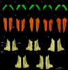
Micro‐CT rendered reconstruction of pterygoid (blue) and ossified portion of the ascending process of palatoquadrate (chartreuse), if any, of five species of Pseudohynobius in the following order from left to right: Ps. flavomaculatus (CIB 17342, the holotype), Ps. jinfo (CIB 85290, the holotype), Ps. guizhouensis (LTHC 0711005, the holotype), Ps. kuankuoshuiensis (ZMC 7504023, the holotype) and Ps. shuichengensis (LTHC 9460084, a paratype). (a–e) Right pterygoid and ossified portion of the ascending process of palatoquadrate, if any, in dorsal (upper) and ventral (lower) views. app, ascending process of palatoquadrate; pt, pterygoid
FIGURE 9
FIGURE 9
Micro‐CT rendered reconstruction of vomer and parasphenoid of five species of Pseudohynobius in the following order from left to right: Ps. flavomaculatus (CIB 17342, the holotype), Ps. jinfo (CIB 85290, the holotype), Ps. guizhouensis (LTHC 0711005, the holotype), Ps. kuankuoshuiensis (ZMC 7504023, the holotype) and Ps. shuichengensis (LTHC 9460084, a paratype). (a–e) Left vomer in ventral (upper) and dorsal (lower) views, (f–j) parasphenoid in ventral (upper), dorsal (middle) and left lateral (lower) views. cfpp, cultriform process of parasphenoid; icf, internal carotid foramen; lap, lateral ala of parasphenoid
FIGURE 10
FIGURE 10
Micro‐CT rendered reconstruction of orbitosphenoid of five species of Pseudohynobius in the following order from left to right: Ps. flavomaculatus (CIB 17342, the holotype), Ps. jinfo (CIB 85290, the holotype), Ps. guizhouensis (LTHC 0711005, the holotype), Ps. kuankuoshuiensis (ZMC 7504023, the holotype) and Ps. shuichengensis (LTHC 9460084, a paratype). (a–e) Left orbitosphenoid in lateral (upper) and medial (middle) views, (f–j) orbitosphenoid in dorsal (left) and ventral (right) views. apo, anteroventral process of orbitosphenoid; fop, foramen opticum; obnf, orbitonasal foramen
FIGURE 11
FIGURE 11
Micro‐CT rendered reconstruction of prootic of four species of Pseudohynobius in the following order from left to right: Ps. flavomaculatus (CIB 17342, the holotype), Ps. guizhouensis (LTHC 0711005, the holotype), Ps. kuankuoshuiensis (ZMC 7504023, the holotype) and Ps. shuichengensis (LTHC 9460084, a paratype); and the prootic‐opisthotic‐exoccipital complex of Ps. jinfo (CIB 85290, the holotype). (a–d) Left prootic and (e) left prootic‐opisthotic‐exoccipital complex in the following views from top to bottom: dorsal, ventral, lateral, medial, anterior and posterior. atpp, anterior process of prootic; auf, foramen acustica; bpp, basal process of prootic; enlf, foramen endolymphaticum; ff, foramen faciale; fnov, fenestra ovalis; fpro, foramen prooticum; lpp, lateral process of prootic; pf, foramen palatinum; prlf, foramen perilymphaticum; psa, prominentia semicircularis anterioris; psof, foramen post‐oticum; vpp, ventral process of prootic
FIGURE 12
FIGURE 12
Micro‐CT rendered reconstruction of opisthotic‐exoccipital complex of four species of Pseudohynobius in the following order from left to right: Ps. flavomaculatus (CIB 17342, the holotype), Ps. guizhouensis (LTHC 0711005, the holotype), Ps. kuankuoshuiensis (ZMC 7504023, the holotype) and Ps. shuichengensis (LTHC 9460084, a paratype); and the stapes of five species of Pseudohynobius in the following order from top to bottom: Ps. flavomaculatus (CIB 17342, the holotype), Ps. jinfo (CIB 85290, the holotype), Ps. guizhouensis (LTHC 0711005, the holotype), Ps. kuankuoshuiensis (ZMC 7504023, the holotype) and Ps. shuichengensis (LTHC 9460084, the paratype). (a–d) Left opisthotic‐exoccipital complex in the following views from top to bottom: dorsal, ventral, medial, anterior and posterolateral, (e–i) left stapes in lateral (left) and dorsal (right) views. fnov, fenestra ovalis; ocp, occipital condyle; prlf, foramen perilymphaticum; psp, prominentia semicircularis posterioris; psof, foramen post‐oticum; ts, tectum synoticum
FIGURE 13
FIGURE 13
Micro‐CT rendered reconstruction of lower jaw of five species of Pseudohynobius in the following order from top to bottom: Ps. flavomaculatus (CIB 17342, the holotype), Ps. jinfo (CIB 85290, the holotype), Ps. guizhouensis (LTHC 0711005, the holotype), Ps. kuankuoshuiensis (ZMC 7504023, the holotype) and Ps. shuichengensis (LTHC 9460084, a paratype). Left lower jaw in (a) dorsal, (b) ventral, (c) medial, (d) lateral views. an, angular; anf, angular foramen; den, dentary; idf, inferior dental foramen; mf, mental foramen; pra, prearticular
FIGURE 14
FIGURE 14
Micro‐CT rendered reconstruction of dentary, prearticular and angular of five species of Pseudohynobius in the following order from left to right: Ps. flavomaculatus (CIB 17342, the holotype), Ps. jinfo (CIB 85290, the holotype), Ps. guizhouensis (LTHC 0711005, the holotype), Ps. kuankuoshuiensis (ZMC 7504023, the holotype) and Ps. shuichengensis (LTHC 9460084, a paratype). (a–e) Left dentary in the following views from top to bottom: lateral, medial, dorsal and ventral, (f–j) left prearticular in the following views from top to bottom: lateral, medial, dorsal and ventral, (k–o) left angular in lateral (upper) and medial (lower) views. anf, angular foramen; idf, inferior dental foramen; mf, mental foramen
FIGURE 15
FIGURE 15
Micro‐CT rendered reconstruction of the ossified hyobranchium of the five species of Pseudohynobius in ventral view: (a) Ps. flavomaculatus (CIB 17342, the holotype), (b) Ps. jinfo (CIB 85290, the holotype), (c) Ps. guizhouensis (LTHC 0711005, the holotype), (d) Ps. kuankuoshuiensis (ZMC 7504023, the holotype), (e) Ps. shuichengensis (LTHC 9460084, a paratype). bb, basibranchial; cb, ceratobranchial; hb, hypobranchial

References

    1. AmphibiaTree . (2007) Rhyacotriton variegatus. Digital Morphology. Available at: http://digimorph.org/specimens/Rhyacotriton_variegatus/head/ [Accessed 9th March 2019]
    1. AmphibiaWeb . (2020) Information on amphibian biology and conservation. Berkeley: The University of California; Available at: http://amphibiaweb.org [Accessed 15th April 2020]
    1. Aoyama, F. (1930) Die Entwicklungsgeschichte des Kopfskelettes des Cryptobranchus japonicas . Zeitschrift für Anatomie und Entwicklungsgeschichte, 93, 107–181 [in German].
    1. Bonebrake, J.E. & Brandon, R.A. (1971) Ontogeny of cranial ossification in the small‐mouthed salamander, Ambystoma texanum (Matthes). Journal of Morphology, 133, 189–204. - PubMed
    1. Brainerd, E.L. (2015) Major transformations in vertebrate breathing mechanisms In: Dial K.P., Shubin N.H. & Brainerd E.L. (Eds.) Great transformations in vertebrate evolution. Chicago and London: The University of Chicago Press, pp. 47–61.

Publication types

LinkOut - more resources