

An attention-supervised full-resolution residual network for the segmentation of breast ultrasound images
- PMID: 32964449
- PMCID: PMC7905659
- DOI: 10.1002/mp.14470
An attention-supervised full-resolution residual network for the segmentation of breast ultrasound images
Abstract
Purpose: Breast cancer is the most common cancer among women worldwide. Medical ultrasound imaging is one of the widely applied breast imaging methods for breast tumors. Automatic breast ultrasound (BUS) image segmentation can measure the size of tumors objectively. However, various ultrasound artifacts hinder segmentation. We proposed an attention-supervised full-resolution residual network (ASFRRN) to segment tumors from BUS images.
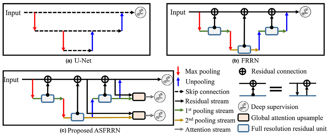
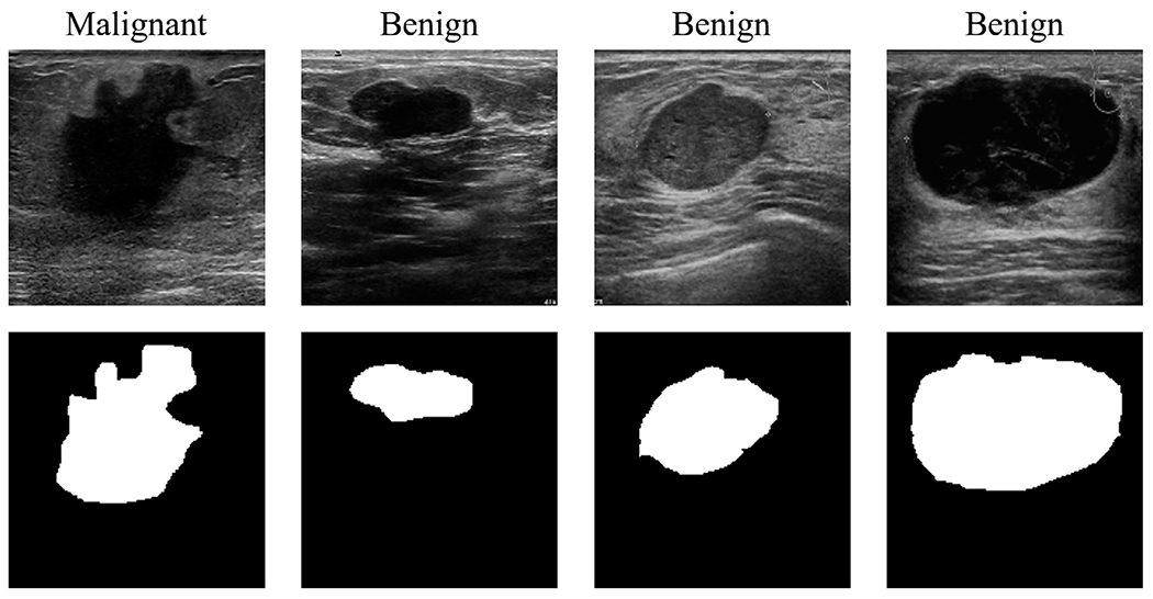
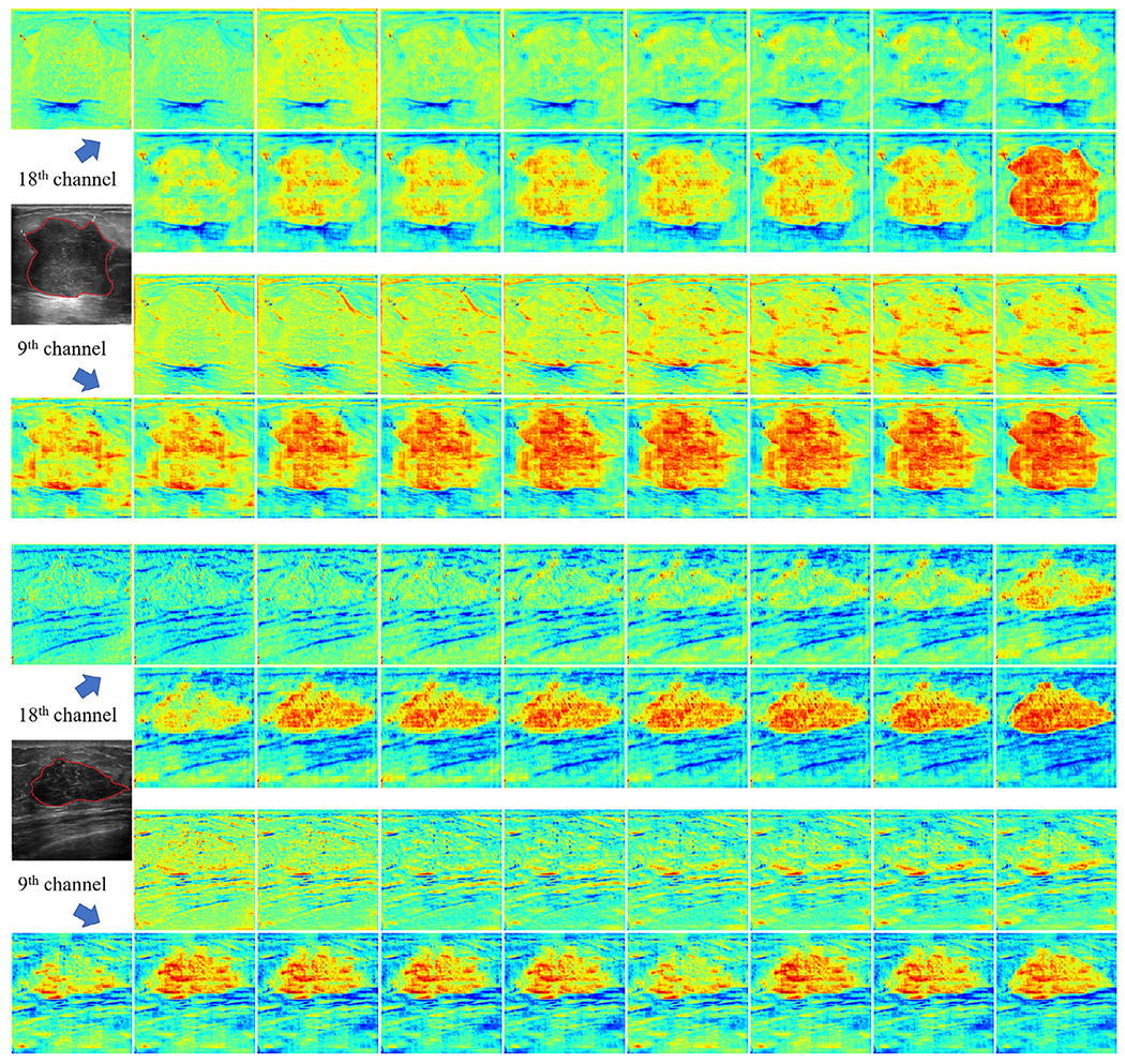
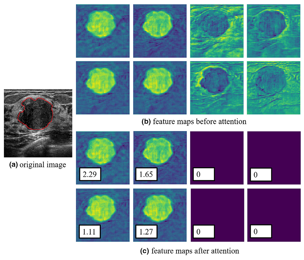
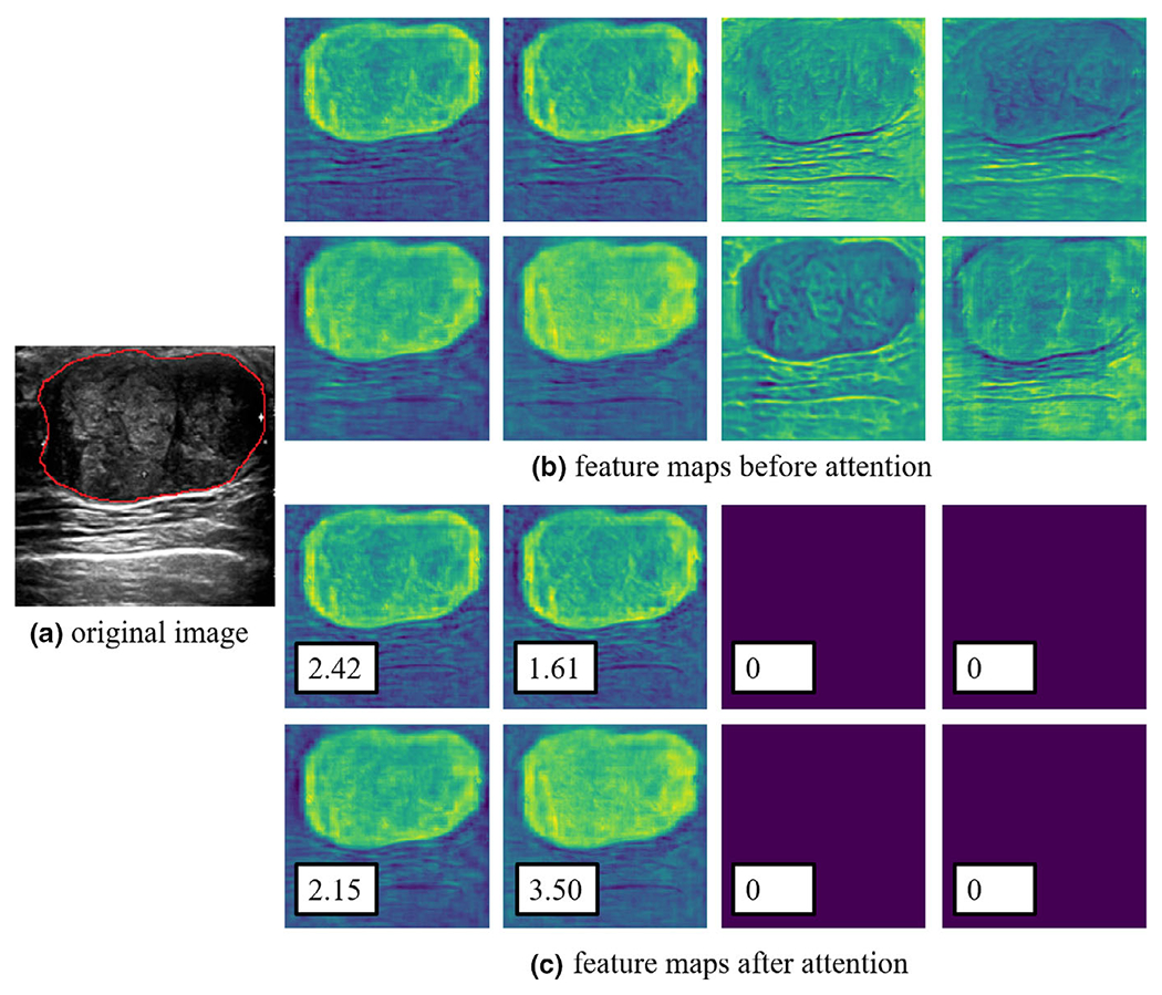
Methods: In the proposed method, Global Attention Upsample (GAU) and deep supervision were introduced into a full-resolution residual network (FRRN), where GAU learns to merge features at different levels with attention for deep supervision. Two datasets were employed for evaluation. One (Dataset A) consisted of 163 BUS images with tumors (53 malignant and 110 benign) from UDIAT Centre Diagnostic, and the other (Dataset B) included 980 BUS images with tumors (595 malignant and 385 benign) from the Sun Yat-sen University Cancer Center. The tumors from both datasets were manually segmented by medical doctors. For evaluation, the Dice coefficient (Dice), Jaccard similarity coefficient (JSC), and F1 score were calculated.
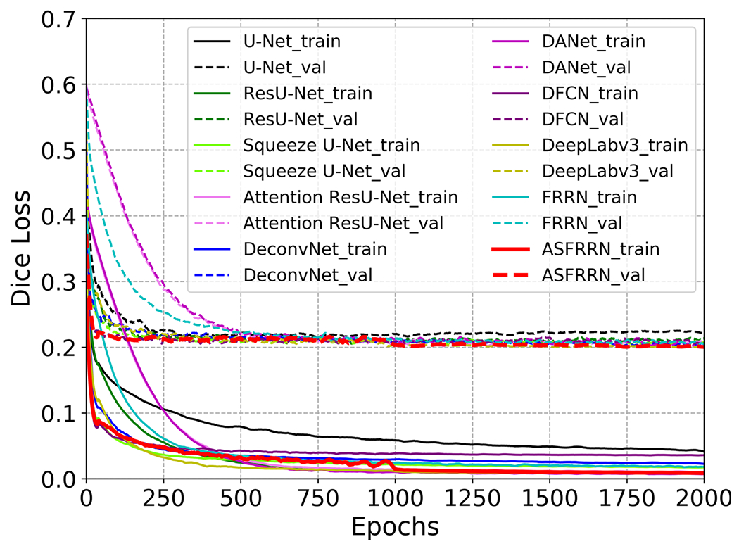
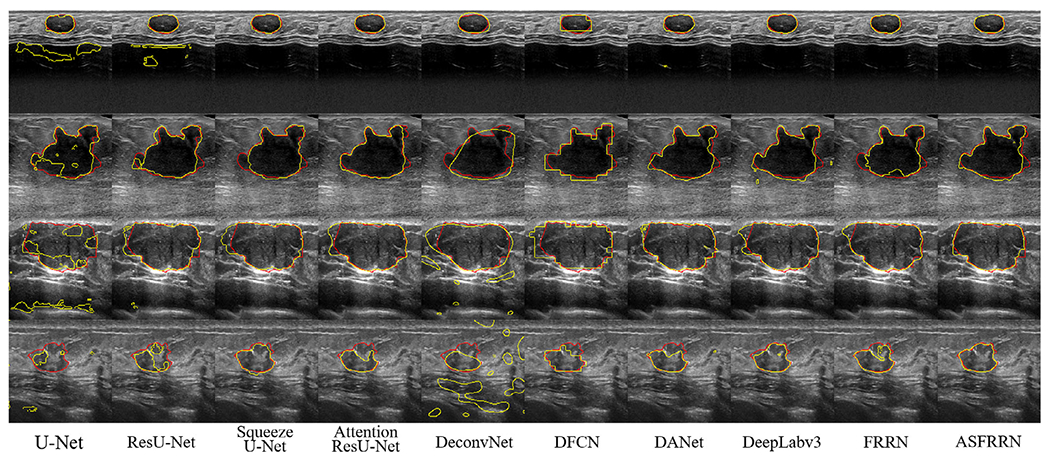
Results: For Dataset A, the proposed method achieved higher Dice (84.3 10.0%), JSC (75.2 10.7%), and F1 score (84.3 10.0%) than the previous best method: FRRN. For Dataset B, the proposed method also achieved higher Dice (90.7 13.0%), JSC (83.7 14.8%), and F1 score (90.7 13.0%) than the previous best methods: DeepLabv3 and dual attention network (DANet). For Dataset A + B, the proposed method achieved higher Dice (90.5 13.1%), JSC (83.3 14.8%), and F1 score (90.5 13.1%) than the previous best method: DeepLabv3. Additionally, the parameter number of ASFRRN was only 10.6 M, which is less than those of DANet (71.4 M) and DeepLabv3 (41.3 M).
Conclusions: We proposed ASFRRN, which combined with FRRN, attention mechanism, and deep supervision to segment tumors from BUS images. It achieved high segmentation accuracy with a reduced parameter number.
Keywords: breast cancer; breast ultrasound image; deep learning; segmentation.
© 2020 American Association of Physicists in Medicine.
Conflict of interest statement
CONFLICT OF INTEREST
The authors have no conflict to disclose.
Figures
Similar articles
-
A deep supervised transformer U-shaped full-resolution residual network for the segmentation of breast ultrasound image.Med Phys. 2023 Dec;50(12):7513-7524. doi: 10.1002/mp.16765. Epub 2023 Oct 10. Med Phys. 2023. PMID: 37816131
-
Comparative Analysis of Current Deep Learning Networks for Breast Lesion Segmentation in Ultrasound Images.Annu Int Conf IEEE Eng Med Biol Soc. 2022 Jul;2022:3878-3881. doi: 10.1109/EMBC48229.2022.9871091. Annu Int Conf IEEE Eng Med Biol Soc. 2022. PMID: 36085645
-
Semi-supervised segmentation of lesion from breast ultrasound images with attentional generative adversarial network.Comput Methods Programs Biomed. 2020 Jun;189:105275. doi: 10.1016/j.cmpb.2019.105275. Epub 2019 Dec 12. Comput Methods Programs Biomed. 2020. PMID: 31978805
-
Breast ultrasound image segmentation: a survey.Int J Comput Assist Radiol Surg. 2017 Mar;12(3):493-507. doi: 10.1007/s11548-016-1513-1. Epub 2017 Jan 9. Int J Comput Assist Radiol Surg. 2017. PMID: 28070777 Review.
-
Methods for the segmentation and classification of breast ultrasound images: a review.J Ultrasound. 2021 Dec;24(4):367-382. doi: 10.1007/s40477-020-00557-5. Epub 2021 Jan 11. J Ultrasound. 2021. PMID: 33428123 Free PMC article. Review.
Cited by
-
Supervised segmentation for guiding peripheral revascularization with forward-viewing, robotically steered ultrasound guidewire.Med Phys. 2023 Jun;50(6):3459-3474. doi: 10.1002/mp.16350. Epub 2023 Mar 21. Med Phys. 2023. PMID: 36906877 Free PMC article.
-
From Images to Genes: Radiogenomics Based on Artificial Intelligence to Achieve Non-Invasive Precision Medicine in Cancer Patients.Adv Sci (Weinh). 2025 Jan;12(2):e2408069. doi: 10.1002/advs.202408069. Epub 2024 Nov 13. Adv Sci (Weinh). 2025. PMID: 39535476 Free PMC article. Review.
References
-
- Siegel RL, Miller KD, Jemal A. Cancer statistics, 2018. CA: Cancer J Clin. 2018;68:7–30. - PubMed
-
- Ohuchi N, Suzuki A, Sobue T, et al. Sensitivity and specificity of mammography and adjunctive ultrasonography to screen for breast cancer in the Japan Strategic Anti-cancer Randomized Trial (J-START) a randomised controlled trial. The Lancet. 2016;387:2381–2382. - PubMed
-
- Di Grezia G, Somma F, Serra N, et al. Reducing costs of breast examination: ultrasound performance and inter-observer variability of expert radiologists versus residents. Cancer Invest. 2016;34:355–360. - PubMed
MeSH terms
Grants and funding
LinkOut - more resources
Full Text Sources
Medical
