

Image Fusion During Standard and Complex Endovascular Aortic Repair, to Fuse or Not to Fuse? A Meta-analysis and Additional Data From a Single-Center Retrospective Cohort
- PMID: 32964768
- PMCID: PMC7816548
- DOI: 10.1177/1526602820960444
Image Fusion During Standard and Complex Endovascular Aortic Repair, to Fuse or Not to Fuse? A Meta-analysis and Additional Data From a Single-Center Retrospective Cohort
Abstract
Purpose: To determine if image fusion will reduce contrast volume, radiation dose, and fluoroscopy and procedure times in standard and complex (fenestrated/branched) endovascular aneurysm repair (EVAR).
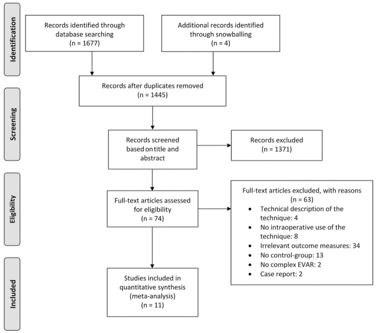
Materials and methods: A search of the PubMed, Embase, and Cochrane databases was performed in December 2019 to identify articles describing results of standard and complex EVAR procedures using image fusion compared with a control group. Study selection, data extraction, and assessment of the methodological quality of the included publications were performed by 2 reviewers working independently. Primary outcomes of the pooled analysis were contrast volume, fluoroscopy time, radiation dose, and procedure time. Eleven articles were identified comprising 1547 patients. Data on 140 patients satisfying the study inclusion criteria were added from the authors' center. Mean differences (MDs) are presented with the 95% confidence interval (CI).
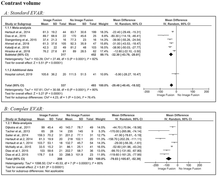
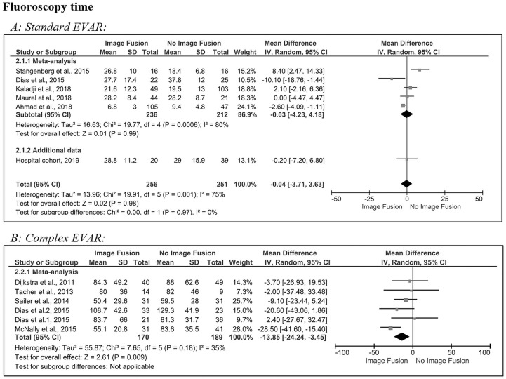
Results: For standard EVAR, contrast volume and procedure time showed a significant reduction with an MD of -29 mL (95% CI -40.5 to -18.5, p<0.001) and -11 minutes (95% CI -21.0 to -1.8, p<0.01), respectively. For complex EVAR, significant reductions in favor of image fusion were found for contrast volume (MD -79 mL, 95% CI -105.7 to -52.4, p<0.001), fluoroscopy time (MD -14 minutes, 95% CI -24.2 to -3.5, p<0.001), and procedure time (MD -52 minutes, 95% CI -75.7 to -27.9, p<0.001).
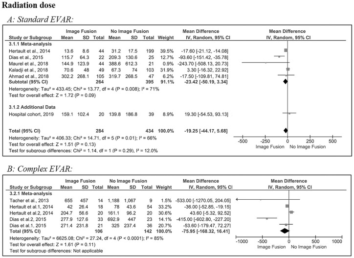
Conclusion: The results of this meta-analysis confirm that image fusion significantly reduces contrast volume, fluoroscopy time, and procedure time in complex EVAR but only contrast volume and procedure time for standard EVAR. Though a reduction was suggested, the radiation dose was not significantly affected by the use of fusion imaging in either standard or complex EVAR.
Keywords: contrast volume; endovascular aneurysm repair; fenestrated/branched EVAR; fluoroscopy time; fusion imaging; image fusion; meta-analysis; procedure time; radiation dose; systematic review.
Conflict of interest statement
Figures

References
-
- Schwein A, Chinnadurai P, Behler G, et al. Computed tomography angiography-fluoroscopy image fusion allows visceral vessel cannulation without angiography during fenestrated endovascular aneurysm repair. J Vasc Surg. 2018;68:2–11. - PubMed
-
- Lindholt JS. Radiocontrast induced nephropathy. Eur J Vasc Endovasc Surg. 2003;25:296–304. - PubMed
-
- Haddad F, Greenberg RK, Walker E, et al. Fenestrated endovascular grafting: the renal side of the story. J Vasc Surg. 2005;41:181–190. - PubMed
-
- Hertault A, Maurel B, Sobocinski J, et al. Impact of hybrid rooms with image fusion on radiation exposure during endovascular aortic repair. Eur J Vasc Endovasc Surg. 2014;48:382–390. - PubMed
-
- Huang IKH, Renani SA, Morgan RA. Complications and reinterventions after fenestrated and branched EVAR in patients with paravisceral and thoracoabdominal aneurysms. Cardiovasc Intervent Radiol. 2018;41:985–997. - PubMed
Publication types
MeSH terms
LinkOut - more resources
Full Text Sources
Research Materials
Miscellaneous
