. 2020 Nov 1;124(5):1388-1398.
doi: 10.1152/jn.00128.2020. Epub 2020 Sep 23.

Cannabidiol activation of vagal afferent neurons requires TRPA1

Affiliations

Cannabidiol activation of vagal afferent neurons requires TRPA1

Cody W Kowalski et al. J Neurophysiol. .

Abstract

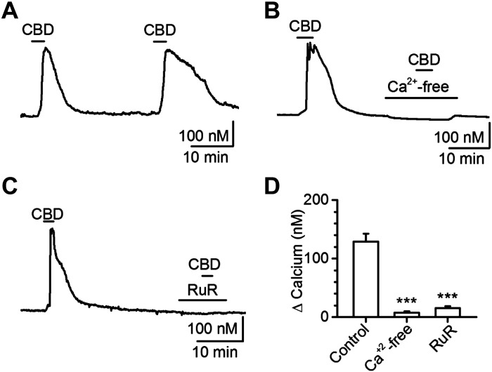
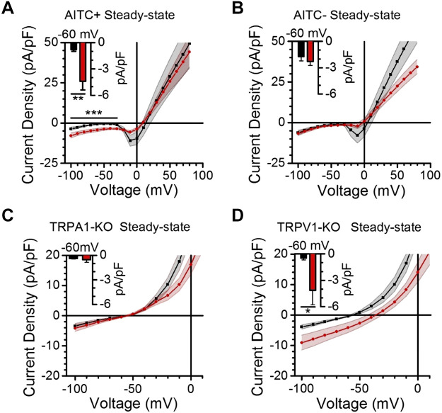
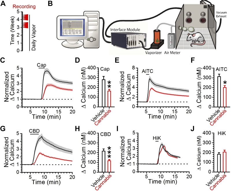
Vagal afferent neurons abundantly express excitatory transient receptor potential (TRP) channels, which strongly influence afferent signaling. Cannabinoids have been identified as direct agonists of TRP channels, including TRPA1 and TRPV1, suggesting that exogenous cannabinoids may influence vagal signaling via TRP channel activation. The diverse therapeutic effects of electrical vagus nerve stimulation also result from administration of the nonpsychotropic cannabinoid, cannabidiol (CBD); however, the direct effects of CBD on vagal afferent signaling remain unknown. We investigated actions of CBD on vagal afferent neurons, using calcium imaging and electrophysiology. CBD produced strong excitatory effects in neurons expressing TRPA1. CBD responses were prevented by removal of bath calcium, ruthenium red, and the TRPA1 antagonist A967079, but not the TRPV1 antagonist SB366791, suggesting an essential role for TRPA1. These pharmacological experiments were confirmed using genetic knockouts where TRPA1 KO mice lacked CBD responses, whereas TRPV1 knockout (KO) mice exhibited CBD-induced activation. We also characterized CBD-provoked inward currents at resting potentials in vagal afferents expressing TRPA1 that were absent in TRPA1 KO mice, but persisted in TRPV1 KO mice. CBD also inhibited voltage-activated sodium conductances in A-fiber, but not in C-fiber afferents. To simulate adaptation, resulting from chronic cannabis use, we administered cannabis extract vapor daily for 3 wk. Cannabis exposure reduced the magnitude of CBD responses, likely due to a loss of TRPA1 signaling. Together, these findings detail a novel excitatory action of CBD at vagal afferent neurons, which requires TRPA1 and may contribute to the vagal mimetic effects of CBD and adaptation following chronic cannabis use.NEW & NOTEWORTHY CBD usage has increased with its legalization. The clinical efficacy of CBD has been demonstrated for conditions including some forms of epilepsy, depression, and anxiety that are also treatable by vagus nerve stimulation. We found CBD exhibited direct excitatory effects on vagal afferent neurons that required TRPA1, were augmented by TRPV1, and attenuated following chronic cannabis vapor exposure. These effects may contribute to vagal mimetic effects of CBD and adaptation after chronic cannabis use.

Keywords: CBD; cannabis; nodose ganglia; transient receptor potential; vagus.


Conflict of interest statement

No conflicts of interest, financial or otherwise, are declared by the authors.

Figures

Fig. 1.
Fig. 1.
Cannabidiol (CBD) provokes depolarization and increases calcium in dissociated vagal afferent neurons. A: cultured vagal afferent neuron cell body with recording pipette in whole cell configuration. B: representative voltage trace of CBD-provoked depolarization and action potential firing. C: quantification of membrane potential at baseline and following CBD-provoked depolarization (n = 13 of 24 neurons/3 rats, ***P < 0.001, paired t test). D: representative images of intracellular calcium concentrations in dissociated vagal afferent neurons under control (left), following bath application of CBD (10 μM, middle), and with potassium-induced depolarization as a positive control (right). Intracellular calcium concentrations are shown with pseudo color by Fura-2 fluorescence ratio according to legend. E: representative calcium trace showing CBD concentration-response relationship in vagal afferents. F: quantification of CBD concentration-response relationship (n = 48 of 144 neurons/6 rats, *P < 0.05, **P < 0.01, and ***P < 0.001, repeated-measures ANOVA with post hoc Holm–Sidak comparison against control). G: population distribution of CBD responsivity.
Fig. 2.
Fig. 2.
Vagal afferent sensitivity to cannabidiol (CBD) is associated with functional expression of TRPA1. A: representative calcium traces of CBD concentration responses from AITC− (left) and from AITC+ (right) vagal afferent neurons. B: average calcium responses across CBD concentrations in TRPA1-lacking (TRPA1−, left) and TRPA1-containing (TRPA1+, right). There was a large difference in CBD responsiveness between TRPA1+ (n = 18 neurons/4 rats) and TRPA1− neurons (n = 19 neurons/4 rats) (P < 0.001, two-way ANOVA). Post hoc comparison against control revealed only the highest concentration of CBD (100 μM) produced a statistically significant increase in calcium in TRPA1– neurons (*P < 0.05); whereas CBD significantly increased calcium concentrations by 1 μM in TRPA1+ neurons (*P < 0.05). Only the highest concentration of CBD (100 μM) produced a statistically significant increase in calcium in TRPA1− neurons. C: population distributions of CBD-nonresponsive and CBD-responsive afferents with regard to their expression of TRPA1 (A1) and TRPV1 (V1) as functionally assayed with AITC (300 μM) and Cap (1 μM). D: scatterplot and linear regression correlating the magnitude of CBD and AITC calcium responses (n = 48 neurons/6 rats, slope = 0.56 ± 0.12, R2 = 0.41, P < 0.0001). AITC, allyl isothiocynate; Cap, capsaicin.
Fig. 3.
Fig. 3.
Vagal afferent cannabidiol (CBD) calcium responses vary according to functional expression of TRPA1 and TRPV1. A: representative calcium trace of CBD responses from a vagal afferent functionally lacking TRPV1 but expressing TRPA1 (TRPV1−/TRPA1+). B: quantification of peak CBD calcium response amplitude from vagal afferents functionally lacking TRPV1 but expressing TRPA1. CBD produced a significant concentration-dependent increase in calcium (n = 8, *P = 0.004, Kruskal–Wallis one-way ANOVA). C: representative calcium trace of CBD responses from a vagal afferent neuron expressing both TRPV1 and TRPA1 (TRPV1+/TRPA1+). D: quantification of peak CBD calcium response amplitude from vagal afferents containing both TRPV1 and TRPA1. There was a main effect of CBD to increase calcium levels across CBD concentrations (n = 21, P < 0.001, Kruskal–Wallis one-way ANOVA) that was qualified by a statistically significant interaction compared with different TRPV1−/TRPA1+ group of neurons (*P = 0.02, two-way ANOVA). AITC, allyl isothiocynate; Cap, capsaicin.
Fig. 4.
Fig. 4.
Cannabidiol (CBD) activation of vagal afferent neurons requires external calcium and is prevented by the nonspecific transient receptor potential (TRP) channel blocker ruthenium red. A: representative calcium trace of 30 μM CBD calcium responses, illustrating lack of desensitization with >25 min washout periods (n = 61 neurons/6 rats, P = 0.64, paired t test). B: representative calcium trace illustrating lack of CBD-calcium response in the absence of extracellular calcium (n = 42 neurons/3 rats, ***P < 0.001, Mann–Whitney rank sum). C: representative calcium trace illustrating lack of CBD-calcium response with the nonspecific TRP-channel pore blocker ruthenium red (RuR; n = 101 neurons/3 rats, ***P < 0.001, Mann–Whitney rank sum). D: quantifications for experiments illustrated in AC.
Fig. 5.
Fig. 5.
Cannabidiol (CBD) activation of vagal afferent neurons requires TRPA1. A: representative calcium trace illustrating a persistent CBD-calcium response with 3 μM of the TRPV1 antagonist. B: representative calcium trace illustrating elimination of the CBD-calcium response with 10 μM of the TRPA1 antagonist A967079. C: quantifications of peak calcium response magnitude from CBD under control conditions and following pretreatment with SB366791 (n = 28 neurons/3 rats, P = 0.36, Mann–Whitney rank sum) or A967079 (n = 87 neurons/4 rats, ***P < 0.001, Mann–Whitney rank sum). D: representative calcium trace illustrating persistent CBD-calcium response in TRPV1 KO. E: representative calcium trace illustrating loss of CBD-calcium response in TRPA1 KO. F: quantification of CBD, AITC, and capsaicin responses from control, TRPV1 KO, and TRPA1 KO mice. Only deletion of TRPA1 prevented CBD-calcium responses (n = 66 neurons/3 mice, ***P < 0.001, Bonferroni corrected t test) as well as AITC-calcium responses as expected (n = 66 neurons/3 mice, ***P < 0.001, Bonferroni corrected t test). AITC, allyl isothiocynate; Cap, capsaicin.
Fig. 6.
Fig. 6.
Cannabidiol (CBD) inhibits voltage-activated sodium channels in capsaicin-insensitive vagal afferent neurons. A: illustration of voltage step protocol used to produce current-voltage curves. B: representative baseline current-voltage traces resulting from step protocol shown in A. Highlighted are the regions used to isolate the steady-state and voltage-activated sodium channel (NaV) component of the trace to generate current-voltage curves, such as the NaV curves in D and E. NaV currents are activated as the neuron is depolarized and not present at very hyperpolarized potentials. C: representative current-voltage traces produced following bath application of 30 μM CBD. D: quantification of NaV current-voltage profiles of dissociated vagal afferents sensitive to capsaicin (Cap). Baseline current-voltage is plotted in black, whereas CBD current-voltage is plotted in red. E: quantification of NaV current-voltage profiles of vagal afferents insensitive to capsaicin. CBD application significantly inhibited NaV selectively in this population (n = 16 neurons/5 rats, P < 0.001, two-way ANOVA; *P < 0.01, Bonferroni’s t tests).
Fig. 7.
Fig. 7.
Cannabidiol (CBD) provokes depolarizing current via TRPA1. A and B: quantifications of steady-state current-voltage profiles of dissociated vagal afferents at baseline in black and following application of 30 μM CBD in red, with the population divided according to sensitivity to the TRPA1 agonist AITC. Current at resting membrane potential (−60 mV) is illustrated with column plots (inset) (n = 16 neurons/8 rats , **P < 0.01, paired t test). CBD provoked significant inward currents at resting potentials, compared with bath control, exclusively in afferents functionally expressing TRPA1 (n = 16 neurons/8 rats, ***P = 0.005, paired t test) but not in those lacking TRPA1 (n = 12 neurons/7 rats, P = 0.14, paired t test). C and D: quantifications of steady-state current-voltage profiles of dissociated vagal afferents, at baseline in black and following application of 30 μM CBD in red. TRPA1 knockout (KO) prevented CBD-provoked inward currents at resting potentials (n = 13 neurons/4 mice, P = 0.36, paired t test) that were evident in TRPV1 KO (n = 15 neurons/3 mice, *P = 0.04, paired t test). AITC, allyl isothiocynate.
Fig. 8.
Fig. 8.
Chronic cannabis vapor exposure attenuates CBD and AITC calcium responses. A: illustration of chronic cannabis vapor administration protocol; cannabis or vehicle vapor was administered for at least 3 wk before tissue collection and recordings (red arrowheads). B: illustration of chronic cannabis vaporization system. C: normalization of vehicle control (n = 27 neurons/4 rats, black trace) and cannabis extract-treated (n = 31 neurons/6 rats, red trace) capsaicin responses. The standard error is represented as a shaded region over each line. D: comparison of peak capsaicin responses in control and cannabis cohorts (**P = 0.002, t test). E: normalization of vehicle control (n = 27 neurons/4 rats, black trace) and cannabis extract-treated (n = 31 neurons/6 rats, red trace) AITC responses. The standard error range is represented as a shaded region over each line. F: comparison of peak AITC calcium responses from control and cannabis vapor-treated cohorts (*P = 0.02, t test). G: normalization of vehicle control (n = 27 neurons/4 rats, black trace) and cannabis extract-treated (n = 31 neurons/6 rats, red trace) CBD responses. The standard error range is represented as a shaded region over each line. H: comparison of peak CBD responses in vehicle control and cannabis extract groups (***P < 0.001, t test). I: normalization of vehicle control (n = 27 neurons/4 rats, black trace) and cannabis extract (n = 31 neurons/6 rats, red trace) to 55 mM potassium (HiK) calcium responses. J: quantification of vehicle control and cannabis extract to 55 mM potassium (HiK) calcium responses (P = 0.47, t test). AITC, allyl isothiocynate; Cap, capsaicin; CBD, cannabidiol.

Similar articles

Cited by

References

    1. Aryana A, Williams MA. Marijuana as a trigger of cardiovascular events: speculation or scientific certainty? Int J Cardiol 118: 141–144, 2007. doi:10.1016/j.ijcard.2006.08.001. - DOI - PubMed
    1. Ashton CH, Moore PB, Gallagher P, Young AH. Cannabinoids in bipolar affective disorder: a review and discussion of their therapeutic potential. J Psychopharmacol 19: 293–300, 2005. doi:10.1177/0269881105051541. - DOI - PubMed
    1. Benowitz NL, Jones RT. Cardiovascular and metabolic considerations in prolonged cannabinoid administration in man. J Clin Pharmacol 21, S1: 214S–223S, 1981. doi:10.1002/j.1552-4604.1981.tb02598.x. - DOI - PubMed
    1. Benowitz NL, Rosenberg J, Rogers W, Bachman J, Jones RT. Cardiovascular effects of intravenous delta-9-tetrahydrocannabinol: autonomic nervous mechanisms. Clin Pharmacol Ther 25: 440–446, 1979. doi:10.1002/cpt1979254440. - DOI - PubMed
    1. Berthoud H-R. The vagus nerve, food intake and obesity. Regul Pept 149: 15–25, 2008. doi:10.1016/j.regpep.2007.08.024. - DOI - PMC - PubMed

Publication types