Review
. 2021 Feb;78(3):889-907.
doi: 10.1007/s00018-020-03641-5. Epub 2020 Sep 23.

Diversity of intrinsically photosensitive retinal ganglion cells: circuits and functions

Affiliations
Review

Diversity of intrinsically photosensitive retinal ganglion cells: circuits and functions

Marcos L Aranda et al. Cell Mol Life Sci. 2021 Feb.

Abstract

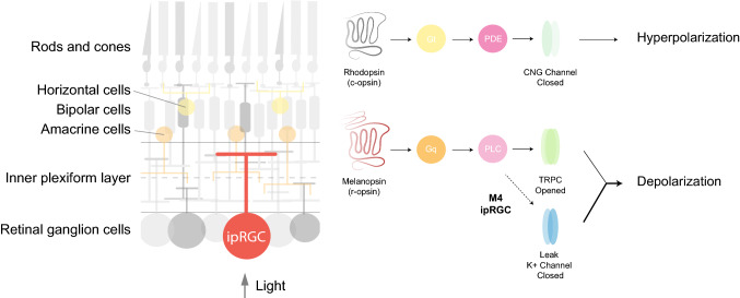
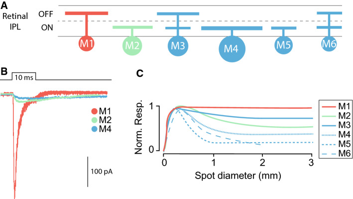
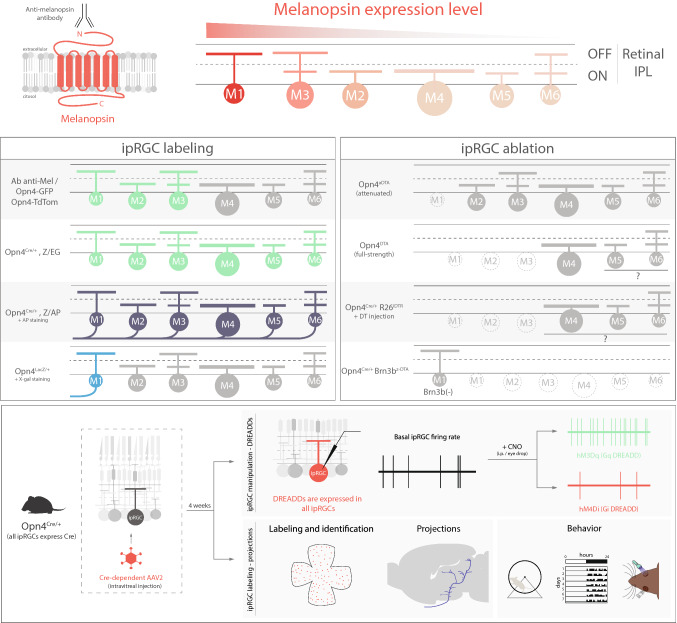
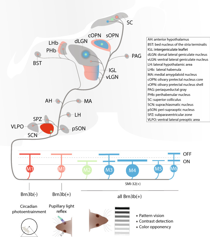
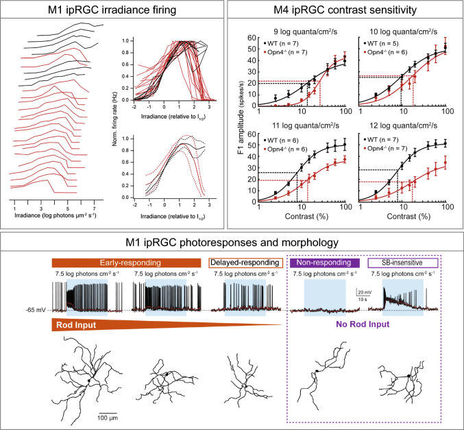
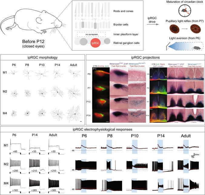
The melanopsin-expressing, intrinsically photosensitive retinal ganglion cells (ipRGCs) are a relatively recently discovered class of atypical ganglion cell photoreceptor. These ipRGCs are a morphologically and physiologically heterogeneous population that project widely throughout the brain and mediate a wide array of visual functions ranging from photoentrainment of our circadian rhythms, to driving the pupillary light reflex to improve visual function, to modulating our mood, alertness, learning, sleep/wakefulness, regulation of body temperature, and even our visual perception. The presence of melanopsin as a unique molecular signature of ipRGCs has allowed for the development of a vast array of molecular and genetic tools to study ipRGC circuits. Given the emerging complexity of this system, this review will provide an overview of the genetic tools and methods used to study ipRGCs, how these tools have been used to dissect their role in a variety of visual circuits and behaviors in mice, and identify important directions for future study.

Keywords: Circadian; Intrinsically photosensitive retinal ganglion cells; Melanopsin; Non-image-forming visual pathway; Pattern vision; Retina.


Figures

Fig. 1
Fig. 1
Simplified schematic representation of a cross-retinal section (left). Signaling cascade of rhodopsin and melanopsin molecules (right). When light actvates rhodopsin, Gt (transducin) triggers an activation of phosphodiesterase (PDE) that closes Na+ channels and induces hyperpolarization. In M1 ipRGCs, melanopsin activates a Gq protein that triggers the activation of phospholipase C (PLC) followed by opening of transient receptor potential channels (TRPC) which give rise to membrane depolarization [24, 92, 101, 136, 137]. In addition to TRPC cascade, in M4 ipRGCs, melanopsin increases cellular excitability via closure of potassium leak channels [55]
Fig. 2
Fig. 2
ipRGC diversity. a Schematic representation of M1–M6 ipRGC stratification in the ON and/or OFF sublaminae of the retinal inner plexiform layer (IPL). b Schematic representation of melanopsin-driven intrinsic electrophysiological responses of M1, M2, and M4 ipRGCs (drawn based on [20]). c Schematic representation of the M1–M6 ipRGCs receptive fields. M1 ipRGC receptive field lacks surround antagonism of the inhibitory input nullifying the surround of the excitatory input. M2–M6 ipRGCs have center–surround-organized receptive fields (drawn based on [9, 10])
Fig. 3
Fig. 3
Diverse toolkit for studying ipRGCs circuits. Top, differential melanopsin expression in M1–M6 ipRGCs. Anti-melanopsin antibody binds to the N-terminus extreme, labeling M1–M3 ipRGC subtypes. ipRGC labeling box: Opn4-GFP line labels M1–M3 ipRGCs in the adult mouse retina and also M4 cells prior to P14 [43]. The Opn4-tdTomato line labels similar subtypes to Opn4-GFP. Opn4Cre/+; Z/EG labels M1–M6 ipRGCs [7]. Opn4Cre/+; Z/AP line labels somas and axons of M1–M6 cells after AP staining [7]. Opn4LacZ/+ line labels M1 ipRGCs somas and axons after X-gal staining [2]. ipRGC ablation box: in the Opn4aDTA line M1 ipRGCs are ablated [19]; in Opn4DTA, M1–M3 cells are likely ablated and Opn4Cre/+R26iDTR mice potentially all ipRGCs are ablated [49, 53]. In Opn4Cre/+Brn3bZ-DTA, all ipRGCs are ablated except Brn3b-negative M1ipRGCs [99]. Bottom, use of adeno-associated viruses (AAV) to manipulate ipRGC activity through DREADDs (top) or to label somata and axons of ipRGCs (bottom) [–, –60]
Fig. 4
Fig. 4
Schematic representation of the main brain targets of ipRGCs. Brn3b-negative M1 ipRGCs are sufficient to drive circadian photoentrainment and project mainly to the SCN, while Brn3b-positive M1 ipRGCs project to sOPN and are necessary for the PLR [99]. Brn3b-positive M1 ipRGCs constitute the majority of sparse M1 ipRGC innervation of the thalamus, hypothalamus, and midbrain (VLPO, SPZ, pSON, AH, MA, vLGN, IGL, dLGN, PAG, BST, PHb, and LHb) [16, 17]. M2 cells project to both image forming (SC and dLGN) and non-image forming (SCN and OPN) visual areas and M4–M6 cells project to brain nuclei involved in image-forming vision such as dorsal geniculate nucleus (dLGN) and superior colliculus (SC) [, –20]. While M3 ipRGCs have been reported to project to the SC [10], no other targets have yet been identified
Fig. 5
Fig. 5
Re-evaluating the roles of ipRGCs. M1 ipRGCs irradiance firing box: every individual M1 cell operates over a narrow range and the population covers irradiances from moonlight to full daylight (left); relations overlaid and aligned by I1/2 for comparison of shape (right) [109]. Melanopsin signaling in both M1 and M4 is active into the scotopic range. M4 ipRGC contrast sensitivity box: contrast response functions of M4 cells in WT (black) and Opn4−/− (red) retinas recorded at background light levels from 9 to 12 log quanta/cm2/s. Vertical dotted lines indicate C50 and horizontal dotted lines indicate half-maximal response [55]. M1 ipRGC photoresponses and morphology box: top, representative recording examples, each from various types of M1 response arranged based on degree of rod input (orange gradient). Blue rectangle indicates start and end of light stimulus. Bottom, dendrite tracing image for each recorded cell [111]
Fig. 6
Fig. 6
ipRGC morphology, projections, and behavior during development. Top, ipRGCs are the only photosensitive cells up to ~ P12, prior to the opening of the eyes and before the connection between bipolar cells and RGCs [115]. ipRGCs set the length of the circadian period [53], and drive rudimentary PLR at P7 [114] and light aversion in mice as young as P6 [138]. ipRGC morphology box: representative cell tracings of M1, M2, and M4 subtypes for P6, P8, P10, P14, and adult time points [44]. ipRGC projections box: left, representative images of olivary pretectal nucleus (OPN) innervation through contralateral (red) and ipsilateral (green) eye injection with fluorescent cholera toxin B subunit (CTB) at P0; Opn4Cre; Z/AP-labeled axons show a similar innervation pattern from P0. In contrast, Opn4LacZ/+-labeled axons are not detected until P7 and show adult-like innervation of the OPN shell (sOPN) by P14 [114]. Right: representative images of suprachiasmatic nucleus (SCN) innervation using CTB. Retinal fibers fill the SCN by P7 and innervation becomes bilateral by P14. Similar innervation pattern was observed in both Opn4Cre; Z/AP and Opn4LacZ/+ lines [114]. ipRGCs electrophysiological responses box: left, representative traces from depolarizing current injection that elicited the maximum spike output for M1, M2, and M4 subtypes at P6, P14, and adult time points [44]. Right, representative light response traces from M1, M2, and M4 cells at P6, P8, P10, P14, and adult time points. Blue rectangle indicates start and end of light stimulus. Black dotted line indicates − 65 mV [44]

References

    1. Berson DM, Dunn FA, Takao M. Phototransduction by retinal ganglion cells that set the circadian clock. Science. 2002;295(5557):1070–1073. doi: 10.1126/science.1067262. - DOI - PubMed
    1. Hattar S, Liao HW, Takao M, Berson DM, Yau KW. Melanopsin-containing retinal ganglion cells: architecture, projections, and intrinsic photosensitivity. Science. 2002;295(5557):1065–1070. doi: 10.1126/science.1069609. - DOI - PMC - PubMed
    1. Altimus CM, Guler AD, Villa KL, McNeill DS, Legates TA, Hattar S. Rods-cones and melanopsin detect light and dark to modulate sleep independent of image formation. Proc Natl Acad Sci USA. 2008;105(50):19998–20003. doi: 10.1073/pnas.0808312105. - DOI - PMC - PubMed
    1. LeGates TA, Fernandez DC, Hattar S. Light as a central modulator of circadian rhythms, sleep and affect. Nat Rev Neurosci. 2014;15(7):443–454. doi: 10.1038/nrn3743. - DOI - PMC - PubMed
    1. Schmidt TM, Alam NM, Chen S, Kofuji P, Li W, Prusky GT, Hattar S. A role for melanopsin in alpha retinal ganglion cells and contrast detection. Neuron. 2014;82(4):781–788. doi: 10.1016/j.neuron.2014.03.022. - DOI - PMC - PubMed

MeSH terms

LinkOut - more resources