. 2020 Dec;37(6):755-765.
doi: 10.1007/s10719-020-09945-9. Epub 2020 Sep 23.

Xylosyltransferase 2 deficiency and organ homeostasis

Affiliations

Xylosyltransferase 2 deficiency and organ homeostasis

Beatrix Ferencz et al. Glycoconj J. 2020 Dec.

Abstract

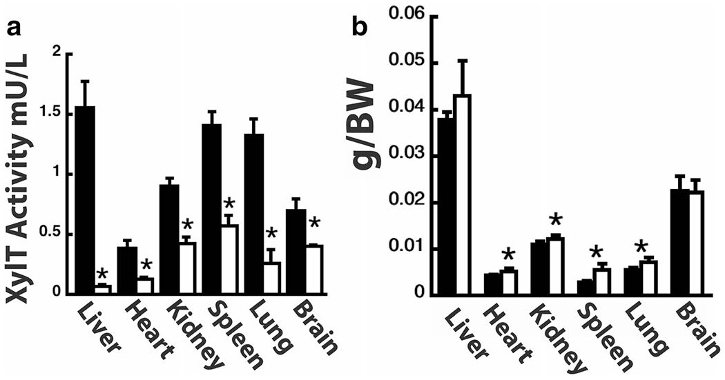
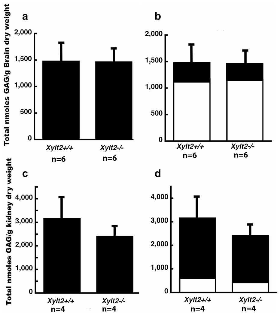
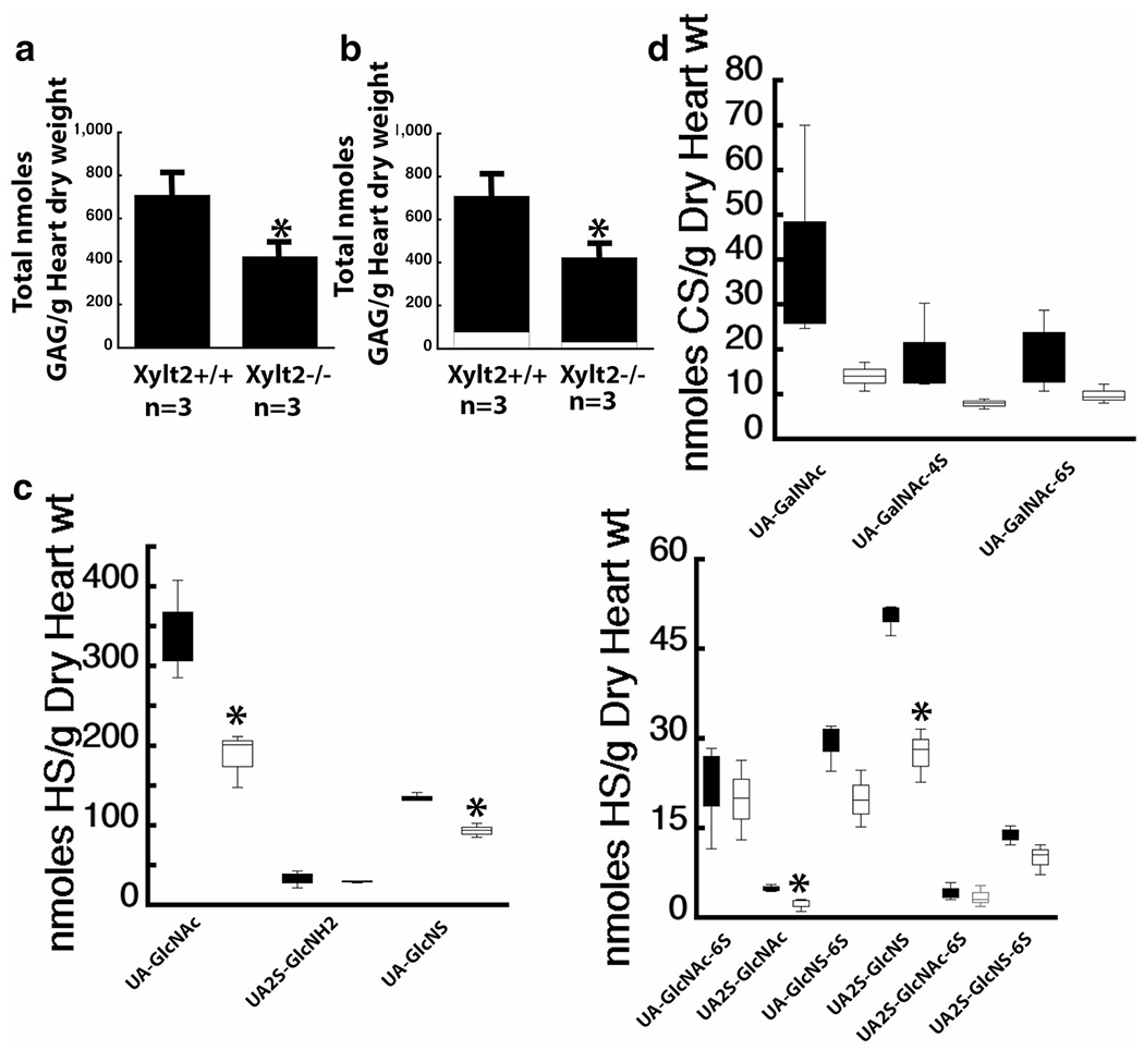
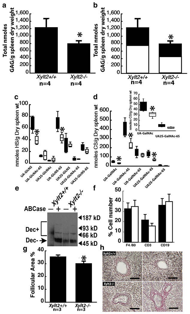
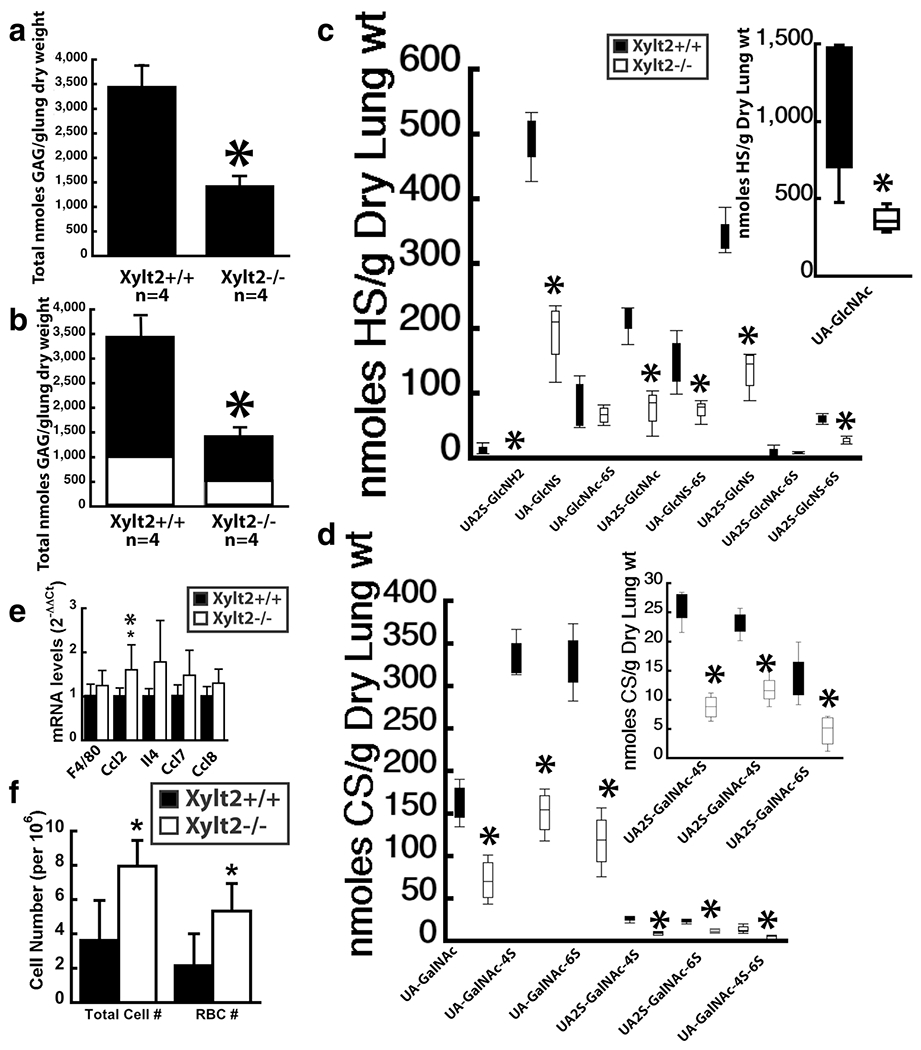
In this paper we characterize the function of Xylosyltransferase 2 (XylT2) in different tissues to investigate the role XylT2 has in the proteoglycan (PG) biochemistry of multiple organs. The results show that in all organs examined there is a widespread and significant decrease in total XylT activity in Xylt2 knock out mice (Xylt2-/-). This decrease results in increased organ weight differences in lung, heart, and spleen. These findings, in addition to our previous findings of increased liver and kidney weight with loss of serum XylT activity, suggest systemic changes in organ function due to loss of XylT2 activity. The Xylt2-/- mice have splenomegaly due to enlargement of the red pulp area and enhanced pulmonary response to bacterial liposaccharide. Tissue glycosaminoglycan composition changes are also found. These results demonstrate a role of XylT2 activity in multiple organs and their PG content. Because the residual XylT activity in the Xylt2-/- is due to xylosyltransferase 1 (XylT1), these studies indicate that both XylT1 and XylT2 have important roles in PG biosynthesis and organ homeostasis.

Keywords: Disaccharide analyses; Extracellular matrix; Genetic modifier; Glycosaminoglycans; Glycotransferase; Organ homeostasis; Proteoglycans; Xylosyltransferase.


Conflict of interest statement

Conflict of interest The authors declare no conflict of interests.

Figures

Fig. 1
Fig. 1
XylT2 deficiency affects multiple organs. a shows XylT activity in organs indicated. n = 5–6 mice, 5–8 month old male for each group. Black Bars Xylt2+/+ mice, Xylt2−/− mice, white bars. (b) Organ weights are changed in Xylt2−/− mice. n = 5 mice for each group, 2–3 month old females. Asterisk indicates significant difference of at least p ≤ 0.05
Fig. 2
Fig. 2
Brain and kidney heparan sulfate and chondroitin sulfate proteoglycans. a shows total brain proteoglycan GAG comparison between Xvlt2+/+ and Xvlt2−/− mice. b shows distribution of HS (dark section) and CS (white section). Error bars are those for total proteoglycans. c shows total proteoglycans in kidney, d shows distribution between HS (black section) and CS (white section). Error bars are those for total proteoglycans
Fig. 3
Fig. 3
XylT2 deficiency significantly decreases HS in heart. a shows heart total GAGs are significantly decreased where the significant decrease is in HS. b shows the distribution of total PGs between HS, black section, and CS, white section. c shows the disaccharide distribution of the significant decreases in Xylt2−/− mice HS, white bars versus the Xylt2+/+, Black bars. Left panel shows high concentration disaccharides and right panel shows low concentration disaccharides. (d) shows the CS disaccharide distribution of Xylt2+/+ mice black bars and Xylt2−/− mice, white bars. Asterisks are significant differences of p ≤ 0.05
Fig. 4
Fig. 4
Xylt2 deficiency results in splenic changes in both HS and CS. a shows a significant decrease in splenic total proteoglycans in the Xylt2−/− mice. b shows that both HS, black section, and CS, white section, are decreased. c shows the HS disaccharide distribution in the spleens. Black bars are Xylt2+/+ mice and white bars are Xylt2−/− mice. d shows the significant decreases in CS disaccharides in the Xylt2−/− spleens. Bars are as in c. Inset is lower concentration disaccharides. e Western blot of splenic protein extracts with decorin antibody. Dec + indicates the smear of decorin core protein with intact CS. Dec-indicates decorin core proteins without CS. Plus and minus signs above the lanes indicate digestion or no digestion with chondroitinase ABC. Lanes 1 and 2 are Xylt2+/+ splenic protein extracts and lanes 3 and 4 are Xylt2−/− splenic extracts. f Fluorescent activated sorting of splenic cells. Bars are as in c. Xylt2+/+, n = 13, Xylt2−/−, n = 4. g Morphometric analyses of splenic follicular area as a percentage to the total splenic area. h Reticular staining of spleens. Upper panels are Xylt2+/+ spleens and lower panels are Xylt2−/− spleens. Scale bars 50 um. Asterisks are significant differences of p ≤ 0.05
Fig. 5
Fig. 5
Lung proteoglycans are significantly altered in the Xylt2−/− mice resulting in inflammatory changes. a shows significant decrease in total lung PGs. b shows both HS (black section) and CS (white section) are significantly decreased. c HS disaccharide distribution in Xylt2+/+ mice (black bars) versus the Xylt2−/− mice (white bars). Inset shows values of UA-GlcNAc levels since they are extremely high. d shows CS disaccharide distribution in the lung. e shows mRNA expression of inflammatory markers in lung, n = 4–6 mice per group (f) Cell counts in bronchiolar lavage fluid from LPS challenged mice, n = 5–6 mice per group. All asterisks are p ≤ 0.05

References

    1. Esko JD, Kimata K, and Lindahl U: Proteoglycans and Sulfated Glycosaminoglycans. In: Essentials of glycobiology. pp. xxix, 784 p. Cold Spring Harbor Laboratory Press, Cold Spring Harbor, N.Y. (2009) - PubMed
    1. Bernfield M, Gotte M, Park PW, Reizes O, Fitzgerald ML, Lincecum J, Zako M: Functions of cell surface heparan sulfate proteoglycans. Annu. Rev. Biochem 68, 729–777 (1999) - PubMed
    1. Esko JD, Selleck SB: Order out of chaos: assembly of ligand binding sites in heparan sulfate. Annu. Rev. Biochem 71, 435–471 (2002) - PubMed
    1. Gustafsson M, Boren J: Mechanism of lipoprotein retention by the extracellular matrix. Curr. Opin. Lipidol 15(5), 505–514 (2004) - PubMed
    1. Mahley RW, Ji ZS: Remnant lipoprotein metabolism: key pathways involving cell-surface heparan sulfate proteoglycans and apolipoprotein E. J. Lipid Res 40(1), 1–16 (1999) - PubMed

Publication types

LinkOut - more resources