

Altered mechanical behavior of demineralized bone following therapeutic radiation
- PMID: 32965711
- PMCID: PMC8212945
- DOI: 10.1002/jor.24868
Altered mechanical behavior of demineralized bone following therapeutic radiation
Abstract
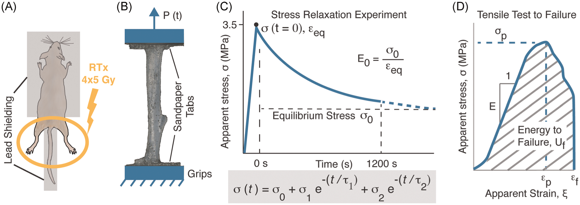
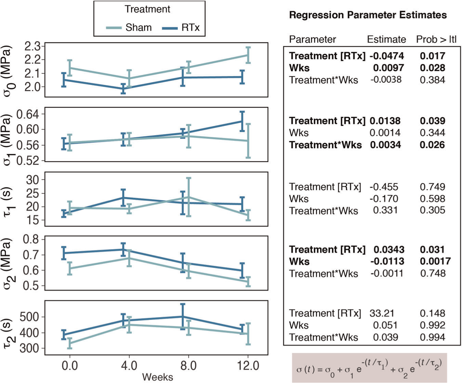
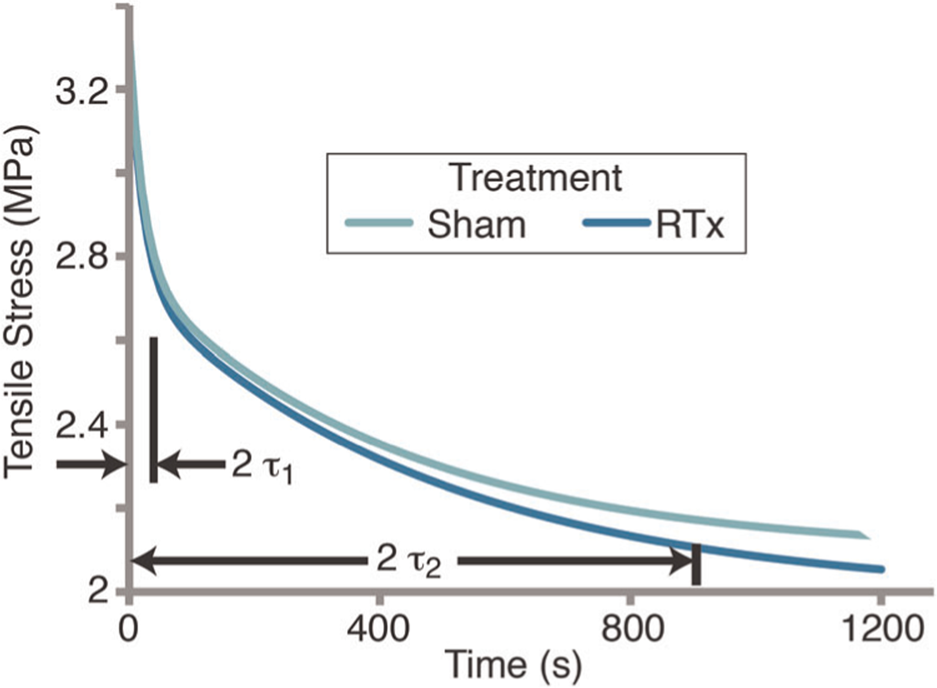
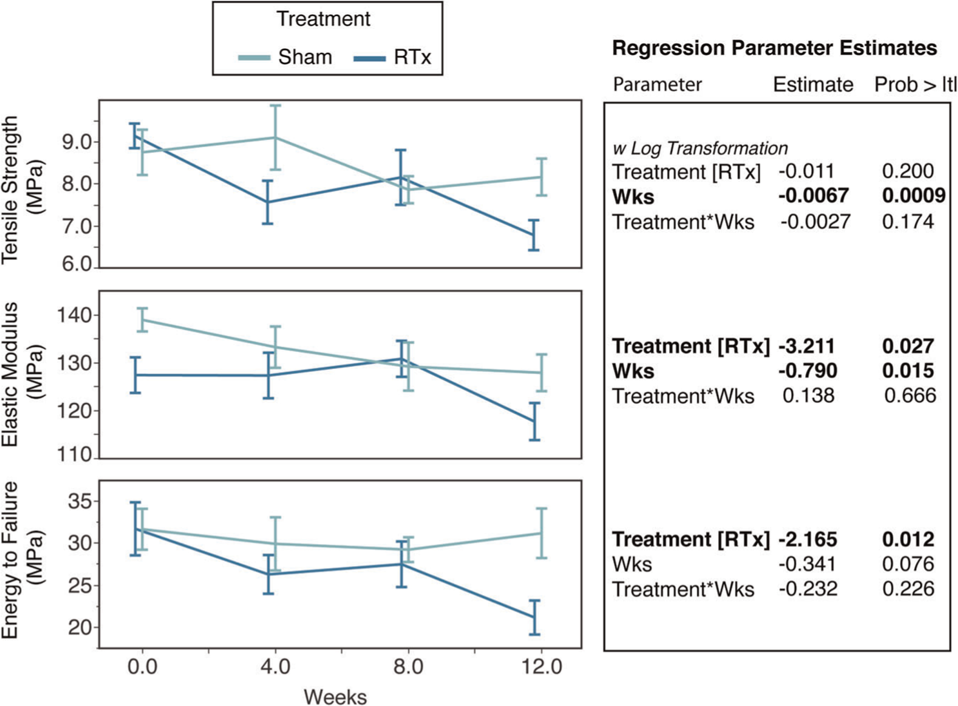
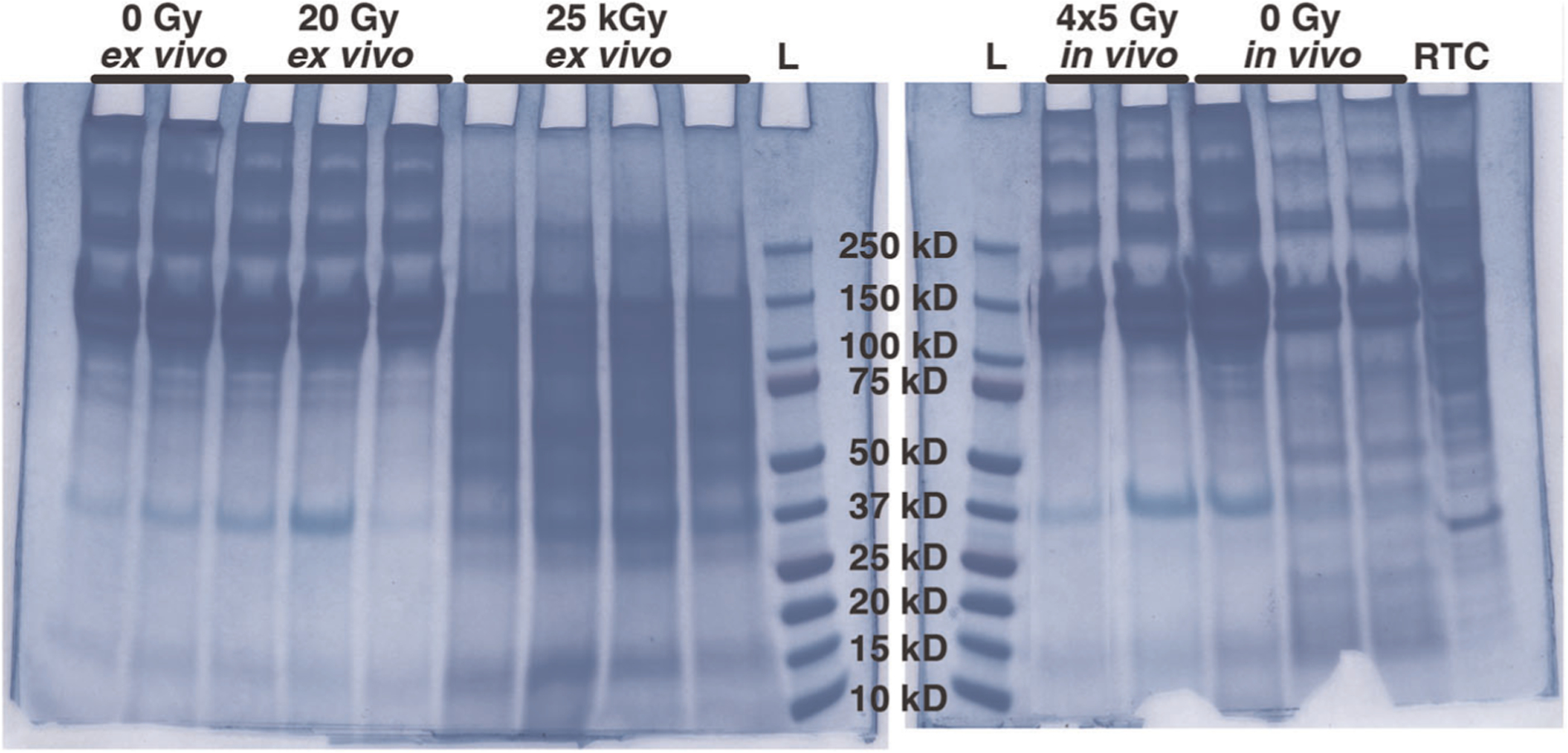
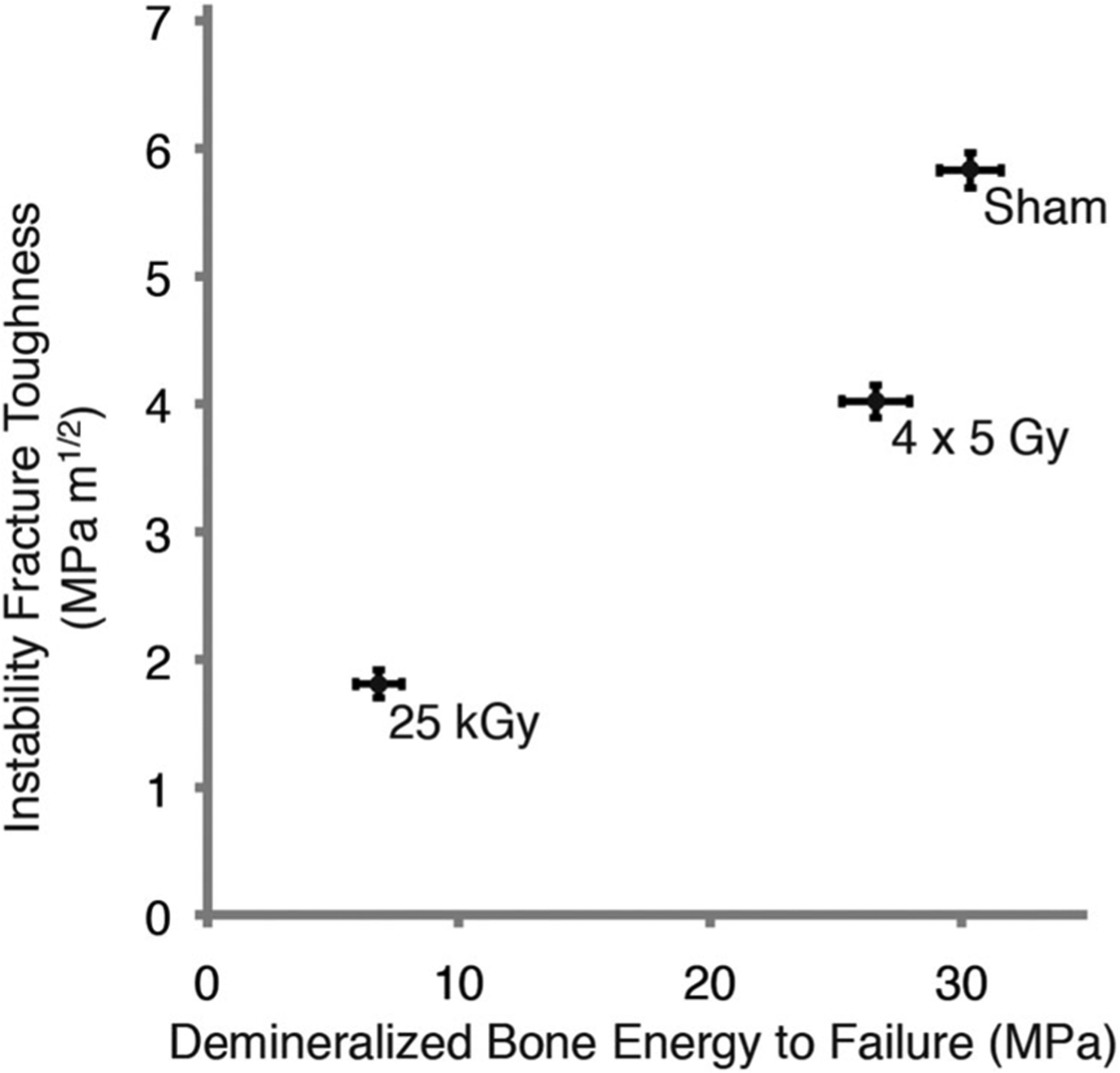
Post-radiotherapy (RTx) bone fragility fractures are a late-onset complication occurring in bone within or underlying the radiation field. These fractures are difficult to predict, as patients do not present with local osteopenia. Using a murine hindlimb RTx model, we previously documented decreased mineralized bone strength and fracture toughness, but alterations in material properties of the organic bone matrix are largely unknown. In this study, 4 days of fractionated hindlimb irradiation (4 × 5 Gy) or Sham irradiation was administered in a mouse model (BALB/cJ, end points: 0, 4, 8, and 12 weeks, n = 15/group/end point). Following demineralization, the viscoelastic stress relaxation, and monotonic tensile mechanical properties of tibiae were determined. Irradiated tibiae demonstrated an immediate (day after last radiation fraction) and sustained (4, 8, 12 weeks) increase in stress relaxation compared to the Sham group, with a 4.4% decrease in equilibrium stress (p < .017). While tensile strength was not different between groups, irradiated tibiae had a lower elastic modulus (-5%, p = .027) and energy to failure (-12.2%, p = .012) with monotonic loading. Gel electrophoresis showed that therapeutic irradiation (4 × 5 Gy) does not result in collagen fragmentation, while irradiation at a common sterilization dose (25 kGy) extensively fragmented collagen. These results suggest that altered collagen mechanical behavior has a role in postirradiation bone fragility, but this can occur without detectable collagen fragmentation. Statement of Clinical Significance: Therapeutic irradiation alters bone organic matrix mechanics and which contribute to diminished fatigue strength, but this does not occur via collagen fragmentation.
Keywords: bone biomechanics; demineralized bone; radiation therapy.
© 2020 Orthopaedic Research Society. Published by Wiley Periodicals LLC.
Figures
Similar articles
-
Limited field radiation therapy results in decreased bone fracture toughness in a murine model.PLoS One. 2018 Oct 3;13(10):e0204928. doi: 10.1371/journal.pone.0204928. eCollection 2018. PLoS One. 2018. PMID: 30281657 Free PMC article.
-
Longitudinal Effects of Single Hindlimb Radiation Therapy on Bone Strength and Morphology at Local and Contralateral Sites.J Bone Miner Res. 2018 Jan;33(1):99-112. doi: 10.1002/jbmr.3289. Epub 2017 Oct 4. J Bone Miner Res. 2018. PMID: 28902435 Free PMC article.
-
Local irradiation alters bone morphology and increases bone fragility in a mouse model.J Biomech. 2010 Oct 19;43(14):2738-46. doi: 10.1016/j.jbiomech.2010.06.017. Epub 2010 Jul 23. J Biomech. 2010. PMID: 20655052
-
Bone embrittlement and collagen modifications due to high-dose gamma-irradiation sterilization.Bone. 2014 Apr;61:71-81. doi: 10.1016/j.bone.2014.01.006. Epub 2014 Jan 16. Bone. 2014. PMID: 24440514
-
Systematic Review on Multilevel Analysis of Radiation Effects on Bone Microarchitecture.Biomed Res Int. 2022 Jun 6;2022:9890633. doi: 10.1155/2022/9890633. eCollection 2022. Biomed Res Int. 2022. PMID: 35782085 Free PMC article.
Cited by
-
Radiation Induces Bone Microenvironment Disruption by Activating the STING-TBK1 Pathway.Medicina (Kaunas). 2023 Jul 16;59(7):1316. doi: 10.3390/medicina59071316. Medicina (Kaunas). 2023. PMID: 37512126 Free PMC article.
-
Fracture Risk of Long Bone Metastases: A Review of Current and New Decision-Making Tools for Prophylactic Surgery.Cancers (Basel). 2021 Jul 21;13(15):3662. doi: 10.3390/cancers13153662. Cancers (Basel). 2021. PMID: 34359563 Free PMC article. Review.
-
Comparison of clinical efficacy of 3D-printed artificial vertebral body and conventional titanium mesh cage in spinal reconstruction after total en bloc spondylectomy for spinal tumors: a systematic review and meta-analysis.Front Oncol. 2024 Feb 6;14:1327319. doi: 10.3389/fonc.2024.1327319. eCollection 2024. Front Oncol. 2024. PMID: 38380368 Free PMC article.
-
Clinical Application of 3D-Printed Artificial Vertebral Body (3DP AVB): A Review.J Pers Med. 2024 Sep 26;14(10):1024. doi: 10.3390/jpm14101024. J Pers Med. 2024. PMID: 39452532 Free PMC article. Review.
-
Orchestrated delivery of PTH [1-34] followed by zoledronic acid prevents radiotherapy-induced bone loss but does not abrogate marrow damage.J Orthop Res. 2022 Dec;40(12):2843-2855. doi: 10.1002/jor.25317. Epub 2022 Mar 25. J Orthop Res. 2022. PMID: 35266584 Free PMC article.
References
-
- Baxter NN. Risk of pelvic fractures in older women following pelvic irradiation. Jama. 2005;294:2587–2593. - PubMed
-
- Dickie CI, Parent AL, Griffin AM, et al. Bone fractures following external beam radiotherapy and limb-preservation surgery for lower extremity soft tissue sarcoma: relationship to irradiated bone length, volume, tumor location and dose. Int J Radiat Oncol Biol Phys. 2009;75: 1119–1124. - PubMed
-
- Gortzak Y, Lockwood GA, Mahendra A, et al. Prediction of pathologic fracture risk of the femur after combined modality treatment of soft tissue sarcoma of the thigh. Cancer. 2010;116:1553–1559. - PubMed
-
- Guise TA. Bone loss and fracture risk associated with cancer therapy. The oncologist. 2006;11:1121–1131. - PubMed
-
- Kwon JW, Huh SJ, Yoon YC, et al. Pelvic bone complications after radiation therapy of uterine cervical cancer: evaluation with MRI. AJR Am J Roentgenol. 2008;191:987–994. - PubMed
Publication types
MeSH terms
Substances
Grants and funding
LinkOut - more resources
Full Text Sources
Medical
